19
views
0
recommends
+1 Recommend
0 collections
    0
    shares
      • Record: found
      • Abstract: found
      • Article: found
      Is Open Access

      Caveolin-1 Associated Adenovirus Entry into Human Corneal Cells

      research-article

      Read this article at

      Bookmark
          There is no author summary for this article yet. Authors can add summaries to their articles on ScienceOpen to make them more accessible to a non-specialist audience.

          Abstract

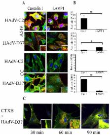
          The cellular entry of viruses represents a critical area of study, not only for viral tropism, but also because viral entry dictates the nature of the immune response elicited upon infection. Epidemic keratoconjunctivitis (EKC), caused by viruses within human adenovirus species D (HAdV-D), is a severe, ocular surface infection associated with corneal inflammation. Clathrin-mediated endocytosis has previously been shown to play a critical role in entry of other HAdV species into many host cell types. However, HAdV-D endocytosis into corneal cells has not been extensively studied. Herein, we show an essential role for cholesterol rich, lipid raft microdomains and caveolin-1, in the entry of HAdV-D37 into primary human corneal fibroblasts. Cholesterol depletion using methyl-β-cyclodextrin (MβCD) profoundly reduced viral infection. When replenished with soluble cholesterol, the effect of MβCD was reversed, allowing productive viral infection. HAdV-D37 DNA was identified in caveolin-1 rich endosomal fractions after infection. Src kinase activity was also increased in caveolin-1 rich endosomal fractions after infection, and Src phosphorylation and CXCL1 induction were both decreased in caveolin-1-/- mice corneas compared to wild type mice. siRNA knock down of caveolin-1 in corneal cells reduced chemokine induction upon viral infection, and caveolin-1-/- mouse corneas showed reduced cellular entry of HAdV-D37. As a control, HAdV-C2, a non-corneal pathogen, appeared to utilize the caveolar pathway for entry into A549 cells, but failed to infect corneal cells entirely, indicating virus and cell specific tropism. Immuno-electron microscopy confirmed the presence of caveolin-1 in HAdV-D37-containing vesicles during the earliest stages of viral entry. Collectively, these experiments indicate for the first time that HAdV-D37 uses a lipid raft mediated caveolin-1 associated pathway for entry into corneal cells, and connects the processes of viral entry with downstream proinflammatory cell signaling.

          Related collections

          Most cited references84

          • Record: found
          • Abstract: found
          • Article: not found

          Isolation of a common receptor for Coxsackie B viruses and adenoviruses 2 and 5.

          A complementary DNA clone has been isolated that encodes a coxsackievirus and adenovirus receptor (CAR). When transfected with CAR complementary DNA, nonpermissive hamster cells became susceptible to coxsackie B virus attachment and infection. Furthermore, consistent with previous studies demonstrating that adenovirus infection depends on attachment of a viral fiber to the target cell, CAR-transfected hamster cells bound adenovirus in a fiber-dependent fashion and showed a 100-fold increase in susceptibility to virus-mediated gene transfer. Identification of CAR as a receptor for these two unrelated and structurally distinct viral pathogens is important for understanding viral pathogenesis and has implications for therapeutic gene delivery with adenovirus vectors.
            Bookmark
            • Record: found
            • Abstract: found
            • Article: not found

            Caveolin, a protein component of caveolae membrane coats.

            Caveolae have been implicated in the transcytosis of macromolecules across endothelial cells and in the receptor-mediated uptake of 5-methyltetrahydrofolate. Structural studies indicate that caveolae are decorated on their cytoplasmic surface by a unique array of filaments or strands that form striated coatings. To understand how these nonclathrin-coated pits function, we performed structural analysis of the striated coat and searched for the molecular component(s) of the coat material. The coat cannot be removed by washing with high salt; however, exposure of membranes to cholesterol-binding drugs caused invaginated caveolae to flatten and the striated coat to disassemble. Antibodies directed against a 22 kd substrate for v-src tyrosine kinase in virus-transformed chick embryo fibroblasts decorated the filaments, suggesting that this molecule is a component of the coat. We have named the molecule caveolin. Caveolae represent a third type of coated membrane specialization that is involved in molecular transport.
              Bookmark
              • Record: found
              • Abstract: found
              • Article: not found

              The multiple faces of caveolae.

              Caveolae are a highly abundant but enigmatic feature of mammalian cells. They form remarkably stable membrane domains at the plasma membrane but can also function as carriers in the exocytic and endocytic pathways. The apparently diverse functions of caveolae, including mechanosensing and lipid regulation, might be linked to their ability to respond to plasma membrane changes, a property that is dependent on their specialized lipid composition and biophysical properties.
                Bookmark

                Author and article information

                Contributors
                Role: Editor
                Journal
                PLoS One
                PLoS ONE
                plos
                plosone
                PLoS ONE
                Public Library of Science (San Francisco, USA )
                1932-6203
                2013
                11 October 2013
                : 8
                : 10
                : e77462
                Affiliations
                [1]Department of Ophthalmology, Harvard Medical School, Massachusetts Eye and Ear Infirmary, Boston, Massachusetts, United States of America
                UC Berkeley, United States of America
                Author notes

                Competing Interests: The authors have declared that no competing interests exist.

                Conceived and designed the experiments: SM AVC JC JR. Performed the experiments: MAY XZ SM AVC JYL MR JR. Analyzed the data: JC JR. Wrote the manuscript: JC JY.

                Article
                PONE-D-13-28911
                10.1371/journal.pone.0077462
                3795695
                24147000
                ed14aa27-8366-4463-877a-4acb71188133
                Copyright @ 2013

                This is an open-access article distributed under the terms of the Creative Commons Attribution License, which permits unrestricted use, distribution, and reproduction in any medium, provided the original author and source are credited.

                History
                : 24 June 2013
                : 9 September 2013
                Funding
                This work was supported by national Institutes of Health (NIH) grants EY013124, EY021558, and EY014104, a Research to Prevent Blindness Senior Scientific Investigator Award (JC), the Falk Foundation, and the Massachusetts Lions Eye Research Fund. The funders had no role in study design, data collection and analysis, decision to publish, or preparation of the manuscript.
                Categories
                Research Article

                Uncategorized
                Uncategorized

                Comments

                Comment on this article