17
views
0
recommends
+1 Recommend
0 collections
    0
    shares
      • Record: found
      • Abstract: found
      • Article: found
      Is Open Access

      Migration and invasion of drug-resistant lung adenocarcinoma cells are dependent on mitochondrial activity

      research-article

      Read this article at

      Bookmark
          There is no author summary for this article yet. Authors can add summaries to their articles on ScienceOpen to make them more accessible to a non-specialist audience.

          Abstract

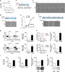
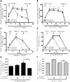
          A small proportion of cancer cells have stem-cell-like properties, are resistant to standard therapy and are associated with a poor prognosis. The metabolism of such drug-resistant cells differs from that of nearby non-resistant cells. In this study, the metabolism of drug-resistant lung adenocarcinoma cells was investigated. The expression of genes associated with oxidative phosphorylation in the mitochondrial membrane was negatively correlated with the prognosis of lung adenocarcinoma. Because the mitochondrial membrane potential (MMP) reflects the functional status of mitochondria and metastasis is the principal cause of death due to cancer, the relationship between MMP and metastasis was evaluated. Cells with a higher MMP exhibited greater migration and invasion than those with a lower MMP. Cells that survived treatment with cisplatin, a standard chemotherapeutic drug for lung adenocarcinoma, exhibited increased MMP and enhanced migration and invasion compared with parental cells. Consistent with these findings, inhibition of mitochondrial activity significantly impeded the migration and invasion of cisplatin-resistant cells. RNA-sequencing analysis indicated that the expression of mitochondrial complex genes was upregulated in cisplatin-resistant cells. These results suggested that drug-resistant cells have a greater MMP and that inhibition of mitochondrial activity could be used to prevent metastasis of drug-resistant lung adenocarcinoma cells.

          Related collections

          Most cited references25

          • Record: found
          • Abstract: found
          • Article: not found

          Evolution of the cancer stem cell model.

          Genetic analyses have shaped much of our understanding of cancer. However, it is becoming increasingly clear that cancer cells display features of normal tissue organization, where cancer stem cells (CSCs) can drive tumor growth. Although often considered as mutually exclusive models to describe tumor heterogeneity, we propose that the genetic and CSC models of cancer can be harmonized by considering the role of genetic diversity and nongenetic influences in contributing to tumor heterogeneity. We offer an approach to integrating CSCs and cancer genetic data that will guide the field in interpreting past observations and designing future studies. Copyright © 2014 Elsevier Inc. All rights reserved.
            Bookmark
            • Record: found
            • Abstract: found
            • Article: found
            Is Open Access

            Metformin inhibits mitochondrial complex I of cancer cells to reduce tumorigenesis

            Recent epidemiological and laboratory-based studies suggest that the anti-diabetic drug metformin prevents cancer progression. How metformin diminishes tumor growth is not fully understood. In this study, we report that in human cancer cells, metformin inhibits mitochondrial complex I (NADH dehydrogenase) activity and cellular respiration. Metformin inhibited cellular proliferation in the presence of glucose, but induced cell death upon glucose deprivation, indicating that cancer cells rely exclusively on glycolysis for survival in the presence of metformin. Metformin also reduced hypoxic activation of hypoxia-inducible factor 1 (HIF-1). All of these effects of metformin were reversed when the metformin-resistant Saccharomyces cerevisiae NADH dehydrogenase NDI1 was overexpressed. In vivo, the administration of metformin to mice inhibited the growth of control human cancer cells but not those expressing NDI1. Thus, we have demonstrated that metformin's inhibitory effects on cancer progression are cancer cell autonomous and depend on its ability to inhibit mitochondrial complex I. DOI: http://dx.doi.org/10.7554/eLife.02242.001
              Bookmark
              • Record: found
              • Abstract: found
              • Article: not found

              A mitochondrial switch promotes tumor metastasis.

              Metastatic progression of cancer is associated with poor outcome, and here we examine metabolic changes underlying this process. Although aerobic glycolysis is known to promote metastasis, we have now identified a different switch primarily affecting mitochondria. The switch involves overload of the electron transport chain (ETC) with preserved mitochondrial functions but increased mitochondrial superoxide production. It provides a metastatic advantage phenocopied by partial ETC inhibition, another situation associated with enhanced superoxide production. Both cases involved protein tyrosine kinases Src and Pyk2 as downstream effectors. Thus, two different events, ETC overload and partial ETC inhibition, promote superoxide-dependent tumor cell migration, invasion, clonogenicity, and metastasis. Consequently, specific scavenging of mitochondrial superoxide with mitoTEMPO blocked tumor cell migration and prevented spontaneous tumor metastasis in murine and human tumor models.
                Bookmark

                Author and article information

                Journal
                Exp Mol Med
                Exp. Mol. Med
                Experimental & Molecular Medicine
                Nature Publishing Group
                1226-3613
                2092-6413
                December 2016
                09 December 2016
                1 December 2016
                : 48
                : 12
                : e277
                Affiliations
                [1 ]Research Institute, National Cancer Center , Goyang, Korea
                [2 ]Department of Molecular Cell Biology, Sungkyunkwan University School of Medicine , Suwon, Korea
                [3 ]Department of System Cancer Science, Graduate School of Cancer Science and Policy, National Cancer Center , Goyang, Korea
                [4 ]Department of Surgery, Yonsei University College of Medicine , Seoul, Korea
                [5 ]Department of Biochemistry and Molecular Biology, Yonsei University College of Medicine , Seoul, Republic of Korea
                Author notes
                [* ]Research Institute, National Cancer Center or Department of System Cancer Science, Graduate School of Cancer Science and Policy, National Cancer Center , Goyang 10408, Korea. E-mail: hjang@ 123456ncc.re.kr
                [6]

                These authors contributed equally to this work.

                Author information
                http://orcid.org/0000-0003-1436-457X
                Article
                emm2016129
                10.1038/emm.2016.129
                5192074
                27932791
                e2243cbf-d268-42c2-a477-777e1acf0ff5
                Copyright © 2016 The Author(s)

                This work is licensed under a Creative Commons Attribution-NonCommercial-ShareAlike 4.0 International License. The images or other third party material in this article are included in the article's Creative Commons license, unless indicated otherwise in the credit line; if the material is not included under the Creative Commons license, users will need to obtain permission from the license holder to reproduce the material. To view a copy of this license, visit http://creativecommons.org/licenses/by-nc-sa/4.0/

                History
                : 02 August 2016
                : 13 September 2016
                : 22 September 2016
                Categories
                Original Article

                Molecular medicine
                Molecular medicine

                Comments

                Comment on this article