14
views
0
recommends
+1 Recommend
0 collections
    0
    shares
      • Record: found
      • Abstract: found
      • Article: found
      Is Open Access

      Farnesyl diphosphate synthase is important for the maintenance of glioblastoma stemness

      research-article

      Read this article at

      Bookmark
          There is no author summary for this article yet. Authors can add summaries to their articles on ScienceOpen to make them more accessible to a non-specialist audience.

          Abstract

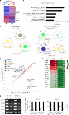
          Glioblastoma is a highly malignant tumor that easily acquires resistance to treatment. The stem-cell-like character (stemness) has been thought to be closely associated with the treatment resistance of glioblastoma cells. In this study, we determined that farnesyl diphosphate synthase (FDPS), a key enzyme in isoprenoid biosynthesis, plays an important role in maintaining glioblastoma stemness. A comparison of the mRNA expression in patient-derived glioblastoma sphere cells, which maintain stemness, and their differentiated counterparts, which lose stemness, via RNA sequencing showed that most of the altered genes were networked in the cholesterol biosynthesis pathway. We screened Federal Drug Administration (FDA)-approved drugs targeting specific enzymes in the cholesterol biosynthesis pathway for their ability to inhibit glioblastoma sphere formation. Inhibitors of FDPS, such as alendronate and zoledronate, significantly reduced the formation of glioblastoma spheres, and alendronate was effective at a lower molar concentration than zoledronate. Knockdown of FDPS using short hairpin RNA also completely inhibited the formation of secondary spheres. FDPS mRNA in patients with glioblastoma was associated with malignancy in three independent microarray data sets. RNA sequencing showed that alendronate treatment reduced the embryonic stem cell signature and activated development- and necrosis-related pathways in glioblastoma spheres. These results suggest that FDPS is important for the maintenance of glioblastoma stemness and that alendronate, a drug widely used to treat osteoporosis, can be repositioned to treat glioblastoma.

          Brain cancer: Enzyme target for potential therapy

          A drug that targets a key enzyme in aggressive brain cancer tumors could help tackle resistance to existing treatments. Glioblastoma is the most aggressive form of brain cancer and remains difficult to treat because the cancer cells can survive chemotherapy and radiotherapy. Certain cells within glioblastoma tumors have ‘stemness’ – unique stem cell-like metabolic characteristics that allow them to rapidly repair DNA damage and trigger relapse. Hyonchol Jang at the National Cancer Center in Goyang, South Korea and co-workers discovered that an enzyme called farnesyl diphosphate synthase (FDPS) helps maintain stemness in glioblastoma. The team then treated patient-derived glioblastoma cells with existing drugs known to inhibit FDPS. One such drug, which is already used to treat osteoporosis, inhibited the formation of secondary glioblastoma and may prove valuble in the treatment of brain cancer.

          Related collections

          Most cited references43

          • Record: found
          • Abstract: found
          • Article: not found

          Evolution of the cancer stem cell model.

          Genetic analyses have shaped much of our understanding of cancer. However, it is becoming increasingly clear that cancer cells display features of normal tissue organization, where cancer stem cells (CSCs) can drive tumor growth. Although often considered as mutually exclusive models to describe tumor heterogeneity, we propose that the genetic and CSC models of cancer can be harmonized by considering the role of genetic diversity and nongenetic influences in contributing to tumor heterogeneity. We offer an approach to integrating CSCs and cancer genetic data that will guide the field in interpreting past observations and designing future studies. Copyright © 2014 Elsevier Inc. All rights reserved.
            Bookmark
            • Record: found
            • Abstract: found
            • Article: not found

            Machine Learning Identifies Stemness Features Associated with Oncogenic Dedifferentiation

            Cancer progression involves the gradual loss of a differentiated phenotype and acquisition of progenitor and stem-cell-like features. Here, we provide novel stemness indices for assessing the degree of oncogenic dedifferentiation. We used an innovative one-class logistic regression (OCLR) machine-learning algorithm to extract transcriptomic and epigenetic feature sets derived from non-transformed pluripotent stem cells and their differentiated progeny. Using OCLR, we were able to identify previously undiscovered biological mechanisms associated with the dedifferentiated oncogenic state. Analyses of the tumor microenvironment revealed unanticipated correlation of cancer stemness with immune checkpoint expression and infiltrating immune cells. We found that the dedifferentiated oncogenic phenotype was generally most prominent in metastatic tumors. Application of our stemness indices to single-cell data revealed patterns of intra-tumor molecular heterogeneity. Finally, the indices allowed for the identification of novel targets and possible targeted therapies aimed at tumor differentiation.
              Bookmark
              • Record: found
              • Abstract: found
              • Article: not found

              A restricted cell population propagates glioblastoma growth following chemotherapy

              Glioblastoma multiforme (GBM) is the most common primary malignant brain tumor, with a median survival of about one year 1 . This poor prognosis is due to therapeutic resistance and tumor recurrence following surgical removal. Precisely how recurrence occurs is unknown. Using a genetically-engineered mouse model of glioma, we identify a subset of endogenous tumor cells that are the source of new tumor cells after the drug, temozolomide (TMZ), is administered to transiently arrest tumor growth. A Nestin-ΔTK-IRES-GFP (Nes-ΔTK-GFP) transgene that labels quiescent subventricular zone adult neural stem cells also labels a subset of endogenous glioma tumor cells. Upon arrest of tumor cell proliferation with TMZ, pulse-chase experiments demonstrate a tumor re-growth cell hierarchy originating with the Nes-ΔTK-GFP transgene subpopulation. Ablation of the GFP+ cells with chronic ganciclovir administration significantly arrested tumor growth and combined TMZ-ganciclovir treatment impeded tumor development. These data indicate the existence of a relatively quiescent subset of endogenous glioma cells that are responsible for sustaining long-term tumor growth through the production of transient populations of highly proliferative cells.
                Bookmark

                Author and article information

                Contributors
                +82-31-920-2239 , hjang@ncc.re.kr
                Journal
                Exp Mol Med
                Exp. Mol. Med
                Experimental & Molecular Medicine
                Nature Publishing Group UK (London )
                1226-3613
                2092-6413
                17 October 2018
                17 October 2018
                October 2018
                : 50
                : 10
                : 137
                Affiliations
                [1 ]ISNI 0000 0004 0628 9810, GRID grid.410914.9, Research Institute, , National Cancer Center, ; Goyang, 10408 Republic of Korea
                [2 ]ISNI 0000 0004 0628 9810, GRID grid.410914.9, Department of Cancer Biomedical Science, , National Cancer Center Graduate School of Cancer Science and Policy, ; Goyang, 10408 Republic of Korea
                [3 ]ISNI 0000 0001 2181 989X, GRID grid.264381.a, Department of Molecular Cell Biology, , Sungkyunkwan University School of Medicine, ; Suwon, 16419 Republic of Korea
                [4 ]ISNI 0000 0004 0628 9810, GRID grid.410914.9, Center for Liver Cancer, , National Cancer Center, ; Goyang, 10408 Republic of Korea
                [5 ]ISNI 0000 0004 0470 5454, GRID grid.15444.30, Department of Neurosurgery, Brain Tumor Center, Severance Hospital, , Yonsei University College of Medicine, ; Seoul, 03722 Korea
                Author information
                http://orcid.org/0000-0001-5676-2037
                http://orcid.org/0000-0003-1436-457X
                Article
                166
                10.1038/s12276-018-0166-2
                6193020
                30333528
                47703a46-e44a-4d26-a682-ebbe7d655493
                © The Author(s) 2018

                Open Access This article is licensed under a Creative Commons Attribution 4.0 International License, which permits use, sharing, adaptation, distribution and reproduction in any medium or format, as long as you give appropriate credit to the original author(s) and the source, provide a link to the Creative Commons license, and indicate if changes were made. The images or other third party material in this article are included in the article’s Creative Commons license, unless indicated otherwise in a credit line to the material. If material is not included in the article’s Creative Commons license and your intended use is not permitted by statutory regulation or exceeds the permitted use, you will need to obtain permission directly from the copyright holder. To view a copy of this license, visit http://creativecommons.org/licenses/by/4.0/.

                History
                : 18 July 2018
                : 4 September 2018
                : 11 September 2018
                Funding
                Funded by: FundRef https://doi.org/10.13039/501100003645, National Cancer Center (NCC);
                Award ID: NCC-1810990
                Award ID: NCC-1810292
                Award ID: NCC-1610190
                Award ID: NCC-1710190
                Award Recipient :
                Funded by: FundRef https://doi.org/10.13039/501100003725, National Research Foundation of Korea (NRF);
                Award ID: NRF-2017R1A2B4009200
                Award Recipient :
                Categories
                Article
                Custom metadata
                © The Author(s) 2018

                Molecular medicine
                Molecular medicine

                Comments

                Comment on this article