14
views
0
recommends
+1 Recommend
0 collections
    0
    shares
      • Record: found
      • Abstract: found
      • Article: found
      Is Open Access

      An evaluation of the stability of image‐quality parameters of Varian on‐board imaging (OBI) and EPID imaging systems

      research-article

      Read this article at

      Bookmark
          There is no author summary for this article yet. Authors can add summaries to their articles on ScienceOpen to make them more accessible to a non-specialist audience.

          Abstract

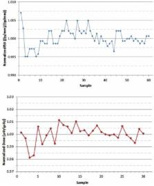
          Quality assurance (QA) of the image quality for image‐guided localization systems is crucial to ensure accurate visualization and localization of regions of interest within the patient. In this study, the temporal stability of selected image parameters was assessed and evaluated for kV CBCT mode, planar radiographic k V, and MV modes. The motivation of the study was to better characterize the temporal variability in specific image‐quality parameters. The CATPHAN, QckV‐1, and QC‐3 phantoms were used to evaluate the image‐quality parameters of the imaging systems on a Varian Novalis Tx linear accelerator. The planar radiographic images were analyzed in PIPSpro with high‐contrast spatial resolution ( f 30 , f 40 , f 50 lp / mm ) being recorded. For OBI kV CBCT, high‐quality head full‐fan acquisition and pelvis half‐fan acquisition modes were evaluated for uniformity, noise, spatial resolution, HU constancy, and geometric distortion. Dose and X‐ray energy for the OBI were recorded using the Unfors RaySafe Xi system with the R/F High Detector for kV planar radiographic and the CT detector for kV CBCT. Dose for the MV EPID was recorded using a PTW975 Semiflex ion chamber, PTW UNIDOS electrometer, and CNMC Plastic Water. For each image‐quality parameter, values were normalized to the mean, and the normalized standard deviations were recorded to evaluate the parameter's temporal variability. For planar radiographic modes, the normalized standard deviations of the spatial resolution ( f 30 , f 40 , & f 50 ) were 0.015, 0.008, 0.004 lp/mm and 0.006, 0.009, 0.018 lp/mm for the kV and MV, respectively. The normalized standard deviation of dose for kV and MV were 0.010 mGy and 0.005 mGy, respectively. The standard deviations for full‐and half‐fan kV CBCT modes were averaged together. The following normalized standard deviations for each kV CBCT parameter were: 0.075 HU (uniformity), 0.071 HU (noise), 0.006 mm (AP‐geometric distortion), 0.005 mm (LAT‐geometric distortion), 0.058 mm (slice thickness), 0.124 (f50), 0.031 (HU constancy – Lung), 0.063 (HU constancy – Water), 0.020 (HU constancy – Bone), 0.006 mGy (Dose – Center), 0.004 mGy (Dose –Periphery). Using control chart analysis, institutional QA tolerances were reported as warning and action thresholds based on 1σ and 2σ thresholds. A study was performed to characterize the stability of image‐quality parameters recommended by AAPM Task Group‐142 for the Varian OBI and EPID imaging systems. Both imaging systems show consistent imaging and dosimetric properties over the evaluated time frame.

          PACS number: 87.10.‐e

          Related collections

          Most cited references14

          • Record: found
          • Abstract: found
          • Article: not found

          Task Group 142 report: quality assurance of medical accelerators.

          The task group (TG) for quality assurance of medical accelerators was constituted by the American Association of Physicists in Medicine's Science Council under the direction of the Radiation Therapy Committee and the Quality Assurance and Outcome Improvement Subcommittee. The task group (TG-142) had two main charges. First to update, as needed, recommendations of Table II of the AAPM TG-40 report on quality assurance and second, to add recommendations for asymmetric jaws, multileaf collimation (MLC), and dynamic/virtual wedges. The TG accomplished the update to TG-40, specifying new test and tolerances, and has added recommendations for not only the new ancillary delivery technologies but also for imaging devices that are part of the linear accelerator. The imaging devices include x-ray imaging, photon portal imaging, and cone-beam CT. The TG report was designed to account for the types of treatments delivered with the particular machine. For example, machines that are used for radiosurgery treatments or intensity-modulated radiotherapy (IMRT) require different tests and/or tolerances. There are specific recommendations for MLC quality assurance for machines performing IMRT. The report also gives recommendations as to action levels for the physicists to implement particular actions, whether they are inspection, scheduled action, or immediate and corrective action. The report is geared to be flexible for the physicist to customize the QA program depending on clinical utility. There are specific tables according to daily, monthly, and annual reviews, along with unique tables for wedge systems, MLC, and imaging checks. The report also gives specific recommendations regarding setup of a QA program by the physicist in regards to building a QA team, establishing procedures, training of personnel, documentation, and end-to-end system checks. The tabulated items of this report have been considerably expanded as compared with the original TG-40 report and the recommended tolerances accommodate differences in the intended use of the machine functionality (non-IMRT, IMRT, and stereotactic delivery).
            Bookmark
            • Record: found
            • Abstract: found
            • Article: not found

            Quality assurance for image-guided radiation therapy utilizing CT-based technologies: a report of the AAPM TG-179.

            Commercial CT-based image-guided radiotherapy (IGRT) systems allow widespread management of geometric variations in patient setup and internal organ motion. This document provides consensus recommendations for quality assurance protocols that ensure patient safety and patient treatment fidelity for such systems.
              Bookmark
              • Record: found
              • Abstract: found
              • Article: not found

              Image guidance for precise conformal radiotherapy.

              To review the state of the art in image-guided precision conformal radiotherapy and to describe how helical tomotherapy compares with the image-guided practices being developed for conventional radiotherapy. Image guidance is beginning to be the fundamental basis for radiotherapy planning, delivery, and verification. Radiotherapy planning requires more precision in the extension and localization of disease. When greater precision is not possible, conformal avoidance methodology may be indicated whereby the margin of disease extension is generous, except where sensitive normal tissues exist. Radiotherapy delivery requires better precision in the definition of treatment volume, on a daily basis if necessary. Helical tomotherapy has been designed to use CT imaging technology to plan, deliver, and verify that the delivery has been carried out as planned. The image-guided processes of helical tomotherapy that enable this goal are described. Examples of the results of helical tomotherapy processes for image-guided intensity-modulated radiotherapy are presented. These processes include megavoltage CT acquisition, automated segmentation of CT images, dose reconstruction using the CT image set, deformable registration of CT images, and reoptimization. Image-guided precision conformal radiotherapy can be used as a tool to treat the tumor yet spare critical structures. Helical tomotherapy has been designed from the ground up as an integrated image-guided intensity-modulated radiotherapy system and allows new verification processes based on megavoltage CT images to be implemented.
                Bookmark

                Author and article information

                Contributors
                gutierrezan@uthscsa.edu
                Journal
                J Appl Clin Med Phys
                J Appl Clin Med Phys
                10.1002/(ISSN)1526-9914
                ACM2
                Journal of Applied Clinical Medical Physics
                John Wiley and Sons Inc. (Hoboken )
                1526-9914
                08 March 2015
                March 2015
                : 16
                : 2 ( doiID: 10.1002/acm2.2015.16.issue-2 )
                : 87-98
                Affiliations
                [ 1 ] Department of Radiation Oncology The University of Texas Health Science Center, San Antonio San Antonio TX USA
                Author notes
                [*] [* ] a Corresponding author: Alonso N. Gutiérrez, Department of Radiation Oncology (G237), University of Texas Health Science Center San Antonio, 7979 Wurzbach Rd, MC 7889, San Antonio, TX 78229, USA; phone: (210) 450 1023; fax: (210) 450 1076; email: gutierrezan@ 123456uthscsa.edu

                Article
                ACM20087
                10.1120/jacmp.v16i2.5088
                5690094
                26103178
                a58cd873-038c-43dd-8cc6-c82df2839c20
                © 2015 The Authors.

                This is an open access article under the terms of the Creative Commons Attribution License, which permits use, distribution and reproduction in any medium, provided the original work is properly cited.

                History
                : 05 May 2014
                : 03 November 2014
                Page count
                Figures: 5, Tables: 7, References: 19, Pages: 12, Words: 5021
                Categories
                Radiation Oncology Physics
                Radiation Oncology Physics
                Custom metadata
                2.0
                acm20087
                March 2015
                Converter:WILEY_ML3GV2_TO_NLMPMC version:5.2.5 mode:remove_FC converted:16.11.2017

                igrt,quality assurance,obi,epid
                igrt, quality assurance, obi, epid

                Comments

                Comment on this article