20
views
0
recommends
+1 Recommend
1 collections
    0
    shares
      • Record: found
      • Abstract: found
      • Article: found
      Is Open Access

      Coinfection with SARS-CoV-2 and Influenza A Virus Increases Disease Severity and Impairs Neutralizing Antibody and CD4 + T Cell Responses

      research-article

      Read this article at

      Bookmark
          There is no author summary for this article yet. Authors can add summaries to their articles on ScienceOpen to make them more accessible to a non-specialist audience.

          ABSTRACT

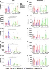
          Given the current coronavirus disease 2019 (COVID-19) pandemic, coinfection of severe acute respiratory syndrome coronavirus 2 (SARS-CoV-2) and influenza A virus (IAV) is a major concern for public health. However, the immunopathogenic events occurring with coinfections of SARS-CoV-2 and IAV remain unclear. Here, we report the pathogenic and immunological consequences of SARS-CoV-2 and IAV H1N1 coinfection in the K18-hACE2 transgenic mouse model. Compared with a single infection with SARS-CoV-2 or IAV, coinfections not only prolonged the primary virus infection period but also increased immune cell infiltration and inflammatory cytokine levels in bronchoalveolar lavage fluid leading to severe pneumonia and lung damage. Moreover, coinfections caused severe lymphopenia in peripheral blood, resulting in reduced total IgG, neutralizing antibody titers, and CD4 + T cell responses against each virus. This study sheds light on the immunopathogenesis of SARS-CoV-2 and IAV coinfection, which may guide the development of effective therapeutic strategies for the treatment of patients coinfected with these viruses.

          IMPORTANCE The cocirculation of influenza virus merging with the COVID-19 pandemic raises a potentially severe threat to public health. Recently, increasing numbers of SARS-CoV-2 and influenza virus coinfection have been reported from many countries. It is a worrisome issue that SARS-CoV-2 coinfection with other pathogens may worsen the clinical outcome and severity of COVID-19 and increase fatality. Here, we evaluated SARS-CoV-2 and IAV coinfection using the K18-hACE2 mouse model. Coinfected mice exhibited increased mortality with prolonged IAV shedding. Furthermore, coinfected mice showed a higher level of cytokines and chemokines than a single infection condition. Interestingly, our data show that coinfected mice showed significantly fewer virus-specific and neutralizing antibodies than the mice with a single infection. Overall, this study suggests that coinfection aggravates viral pathology by impaired neutralizing antibody response.

          Related collections

          Most cited references38

          • Record: found
          • Abstract: found
          • Article: not found

          Clinical features of patients infected with 2019 novel coronavirus in Wuhan, China

          Summary Background A recent cluster of pneumonia cases in Wuhan, China, was caused by a novel betacoronavirus, the 2019 novel coronavirus (2019-nCoV). We report the epidemiological, clinical, laboratory, and radiological characteristics and treatment and clinical outcomes of these patients. Methods All patients with suspected 2019-nCoV were admitted to a designated hospital in Wuhan. We prospectively collected and analysed data on patients with laboratory-confirmed 2019-nCoV infection by real-time RT-PCR and next-generation sequencing. Data were obtained with standardised data collection forms shared by WHO and the International Severe Acute Respiratory and Emerging Infection Consortium from electronic medical records. Researchers also directly communicated with patients or their families to ascertain epidemiological and symptom data. Outcomes were also compared between patients who had been admitted to the intensive care unit (ICU) and those who had not. Findings By Jan 2, 2020, 41 admitted hospital patients had been identified as having laboratory-confirmed 2019-nCoV infection. Most of the infected patients were men (30 [73%] of 41); less than half had underlying diseases (13 [32%]), including diabetes (eight [20%]), hypertension (six [15%]), and cardiovascular disease (six [15%]). Median age was 49·0 years (IQR 41·0–58·0). 27 (66%) of 41 patients had been exposed to Huanan seafood market. One family cluster was found. Common symptoms at onset of illness were fever (40 [98%] of 41 patients), cough (31 [76%]), and myalgia or fatigue (18 [44%]); less common symptoms were sputum production (11 [28%] of 39), headache (three [8%] of 38), haemoptysis (two [5%] of 39), and diarrhoea (one [3%] of 38). Dyspnoea developed in 22 (55%) of 40 patients (median time from illness onset to dyspnoea 8·0 days [IQR 5·0–13·0]). 26 (63%) of 41 patients had lymphopenia. All 41 patients had pneumonia with abnormal findings on chest CT. Complications included acute respiratory distress syndrome (12 [29%]), RNAaemia (six [15%]), acute cardiac injury (five [12%]) and secondary infection (four [10%]). 13 (32%) patients were admitted to an ICU and six (15%) died. Compared with non-ICU patients, ICU patients had higher plasma levels of IL2, IL7, IL10, GSCF, IP10, MCP1, MIP1A, and TNFα. Interpretation The 2019-nCoV infection caused clusters of severe respiratory illness similar to severe acute respiratory syndrome coronavirus and was associated with ICU admission and high mortality. Major gaps in our knowledge of the origin, epidemiology, duration of human transmission, and clinical spectrum of disease need fulfilment by future studies. Funding Ministry of Science and Technology, Chinese Academy of Medical Sciences, National Natural Science Foundation of China, and Beijing Municipal Science and Technology Commission.
            Bookmark
            • Record: found
            • Abstract: found
            • Article: not found

            Clinical Characteristics of Coronavirus Disease 2019 in China

            Abstract Background Since December 2019, when coronavirus disease 2019 (Covid-19) emerged in Wuhan city and rapidly spread throughout China, data have been needed on the clinical characteristics of the affected patients. Methods We extracted data regarding 1099 patients with laboratory-confirmed Covid-19 from 552 hospitals in 30 provinces, autonomous regions, and municipalities in mainland China through January 29, 2020. The primary composite end point was admission to an intensive care unit (ICU), the use of mechanical ventilation, or death. Results The median age of the patients was 47 years; 41.9% of the patients were female. The primary composite end point occurred in 67 patients (6.1%), including 5.0% who were admitted to the ICU, 2.3% who underwent invasive mechanical ventilation, and 1.4% who died. Only 1.9% of the patients had a history of direct contact with wildlife. Among nonresidents of Wuhan, 72.3% had contact with residents of Wuhan, including 31.3% who had visited the city. The most common symptoms were fever (43.8% on admission and 88.7% during hospitalization) and cough (67.8%). Diarrhea was uncommon (3.8%). The median incubation period was 4 days (interquartile range, 2 to 7). On admission, ground-glass opacity was the most common radiologic finding on chest computed tomography (CT) (56.4%). No radiographic or CT abnormality was found in 157 of 877 patients (17.9%) with nonsevere disease and in 5 of 173 patients (2.9%) with severe disease. Lymphocytopenia was present in 83.2% of the patients on admission. Conclusions During the first 2 months of the current outbreak, Covid-19 spread rapidly throughout China and caused varying degrees of illness. Patients often presented without fever, and many did not have abnormal radiologic findings. (Funded by the National Health Commission of China and others.)
              Bookmark
              • Record: found
              • Abstract: found
              • Article: not found

              Clinical and immunologic features in severe and moderate Coronavirus Disease 2019

              Journal of Clinical Investigation
                Bookmark

                Author and article information

                Contributors
                Role: Editor
                Journal
                J Virol
                J Virol
                JVI
                Journal of Virology
                American Society for Microbiology (1752 N St., N.W., Washington, DC )
                0022-538X
                1098-5514
                2 February 2022
                23 March 2022
                March 2022
                23 March 2022
                : 96
                : 6
                : e01873-21
                Affiliations
                [a ] College of Medicine and Medical Research Institute, Chungbuk National Universitygrid.254229.a, , Cheongju, Republic of Korea
                [b ] Infectious Disease Research Center, Korea Research Institute of Bioscience and Biotechnology, University of Science and Technology, Daejeon, Republic of Korea
                [c ] Department of Cancer Biology and Global Center for Pathogens Research and Human Health, Lerner Research Institute, Cleveland Clinic, Cleveland, Ohio, USA
                [d ] Center for Study of Emerging and Re-emerging Viruses, Korea Virus Research Institute, Institute for Basic Science (IBS), Daejeon, Republic of Korea
                [e ] Vinmec Research Institute of Stem Cell and Gene Technology, Vinmec Healthcare System, Hanoi, Vietnam
                Emory University School of Medicine
                Author notes

                Eun-Ha Kim and Thi-Quyen Nguyen contributed equally to this article. Author order was determined by the corresponding author after negotiation.

                The authors declare no conflict of interest.

                Author information
                https://orcid.org/0000-0003-4559-8774
                https://orcid.org/0000-0002-0872-0147
                Article
                01873-21 jvi.01873-21
                10.1128/jvi.01873-21
                8941868
                35107382
                4ba03da6-0a44-4856-93b8-746ea83fa838
                Copyright © 2022 Kim et al.

                This is an open-access article distributed under the terms of the Creative Commons Attribution 4.0 International license.

                History
                : 29 October 2021
                : 14 January 2022
                Page count
                Figures: 5, Tables: 0, Equations: 0, References: 40, Pages: 15, Words: 8414
                Funding
                Funded by: Institute for Basic Science (IBS), FundRef https://doi.org/10.13039/501100010446;
                Award ID: IBS-R801-D1
                Award Recipient :
                Funded by: Korea Research Institute of Bioscience and Biotechnology (KRIBB), FundRef https://doi.org/10.13039/501100003715;
                Award ID: KGM9942011
                Award Recipient :
                Funded by: MOHW | Korea National Institute of Health (KNIH), FundRef https://doi.org/10.13039/501100003653;
                Award ID: AI140705
                Award ID: AI140705S1
                Award ID: AI152190
                Award Recipient :
                Categories
                Pathogenesis and Immunity
                virology, Virology
                Custom metadata
                March 2022

                Microbiology & Virology
                sars-cov-2,influenza a virus,coinfection,hace2 mice,neutralizing antibody,t cell,cytokine,chemokine,bronchoalveolar lavage fluid,immune response

                Comments

                Comment on this article