3
views
0
recommends
+1 Recommend
0 collections
    0
    shares
      • Record: found
      • Abstract: found
      • Article: found
      Is Open Access

      Application of the direct in-scope suction technique in antegrade flexible ureteroscopic lithotripsy for the removal of a large ureteric calculus in a kidney transplant recipient: A case report

      case-report

      Read this article at

      Bookmark
          There is no author summary for this article yet. Authors can add summaries to their articles on ScienceOpen to make them more accessible to a non-specialist audience.

          Abstract

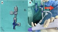
          The occurrence of a large ureteric calculus in a transplanted kidney, originating from a donor, is a rare but significant complication. It poses risks such as urinary obstruction, septicemia, and potential loss of allograft function. In this case, we report our first use of the direct in-scope suction technique during antegrade flexible ureteroscopy lithotripsy. This method successfully removed a donor-derived ureteric calculus in a kidney transplant recipient. The procedure resulted in complete stone removal, and the patient experienced a favorable postoperative recovery without additional adverse events.

          Related collections

          Most cited references15

          • Record: found
          • Abstract: found
          • Article: not found

          Organ shortage crisis: problems and possible solutions.

          G.M Abouna (2008)
          The demand for organ transplantation has rapidly increased all over the world during the past decade due to the increased incidence of vital organ failure, the rising success and greater improvement in posttransplant outcome. However, the unavailability of adequate organs for transplantation to meet the existing demand has resulted in major organ shortage crises. As a result there has been a major increase in the number of patients on transplant waiting lists as well as in the number of patients dying while on the waiting list. In the United States, for example, the number of patients on the waiting list in the year 2006 had risen to over 95,000, while the number of patient deaths was over 6,300. This organ shortage crisis has deprived thousands of patients of a new and better quality of life and has caused a substantial increase in the cost of alternative medical care such as dialysis. There are several procedures and pathways which have been shown to provide practical and effective solutions to this crisis. These include implementation of appropriate educational programs for the public and hospital staff regarding the need and benefits of organ donation, the appropriate utilization of marginal (extended criteria donors), acceptance of paired organ donation, the acceptance of the concept of "presumed consent," implementation of a system of "rewarded gifting" for the family of the diseased donor and also for the living donor, developing an altruistic system of donation from a living donor to an unknown recipient, and accepting the concept of a controlled system of financial payment for the donor. As is outlined in this presentation, we strongly believe that the implementation of these pathways for obtaining organs from the living and the dead donors, with appropriate consideration of the ethical, religious and social criteria of the society, the organ shortage crisis will be eliminated and many lives will be saved through the process of organ donation and transplantation.
            Bookmark
            • Record: found
            • Abstract: found
            • Article: found
            Is Open Access

            Urinary Stones following Renal Transplantation

            Background: The formation of urinary tract stones following renal transplantation is a rare complication. The clinical features of stones after transplantation differ from those of non-transplant patients. Renal colic or pain is usually absent and rarely resembles acute rejection. Methods: We retrospectively studied 849 consecutive kidney transplant patients in The Rogosin Institute/The Weill-Cornell Medical Center, New York who were transplanted between 1980 and 1997 and had functioning grafts for more than 3 months, to determine the incidence of stone formation, composition, risk factors and patient outcome. Results: At our center, urinary stones were diagnosed in 15 patients (1.8%) of 849 functioning renal grafts for 3 or more months. Of the 15 patients, 10 were males and 5 were females in their third and fourth decade. Eight patients received their transplant from living donors and 7 from cadaveric donors. The stones were first diagnosed between 3 and 109 months after transplantation (mean 17.8 months) and 5 patients had recurrent episodes. The stones were located in the bladder in 11 cases (73.3%), transplanted kidney in 3 cases and in multiple sites in one case. The size of stones varied from 3.4 mm to 40 mm (mean 12 mm). The composition of stones was a mixed form of calcium oxalate and calcium phosphate in 5 cases and 4 patients had infected stones consisting of struvite or mixed form of struvite and calcium phosphate. Factors predisposing to stone formation included tertiary hyperparathyroidism (n=8), hypercalciuria (n=5), recurrent urinary tract infection (n=5), hypocitraturia (n=4), and obstructive uropathy (n=2). Many cases had more than one risk factor. Clinically, painless hematuria was observed in 6 patients and dysuria without bacteriuria in 5 patients. None had renal colic or severe pain at any time. There were no changes in graft function at diagnosis and after removal of stones. Five patients passed stones spontaneously and 8 patients underwent cystoscopy for stone removal. Conclusion: Urinary stone formation following kidney transplantation is a rare complication (1.8%). Hyperparathyroidism, hypercalciuria, recurrent urinary tract infection and hypocitraturia are the most common risk factors, but often there are multiple factors which predispose to stone formation. To detect stones and determine their location and size, ultrasonography appears to be the most useful diagnostic tool. Prompt diagnosis, the removal of stones and stone-preventive measures can prevent adverse effects on renal graft outcome.
              Bookmark
              • Record: found
              • Abstract: found
              • Article: found
              Is Open Access

              Implementation of Supine Percutaneous Nephroscopic Surgery to Remove an Upward Migration of Ureteral Catheter in Infancy: A Case Report

              Background Double-J stents are favorably utilized after pyeloplasty. In rare situations, the stent may migrate upward. Here, we demonstrate the implementation and result of a supine percutaneous nephroscopic surgery (PNS) to retrieve a proximately migrated ureteral catheter in a pediatric patient. Patient and Methods A 1-year-old boy was suffering from an upward migration of a ureteric catheter into the right ureter after an open Anderson-Hynes pyeloplasty. The child was placed in the Galdakao-modified supine Valdivia (GMSV) position and a PNS procedure was performed. The calyceal access was carefully punctured by ultrasonographic guidance. The nephrostomy tract was dilated with a metal dilator using a one-step technique. An exploratory nephroscopy of the renal pelvis was conducted with a 12Fr miniature nephroscope and the migrated ureteral catheter was removed. A hybrid guidewire was retrogradely inserted into the ureteric orifice using a rigid ureteroscope. An antegrade double J stent was inserted in the proper position and a percutaneous nephrostomy was performed. Results and Conclusion This is the first report of a successfully removed upwardly migrated ureteral catheter with concurrent insertion of an antegrade double J stent by supine PNS in the GMSV position in an infant. The patient recovered well after surgery with no adverse event, demonstrating that this operation can be carried out safely on pediatric patients.
                Bookmark

                Author and article information

                Contributors
                Journal
                Urol Case Rep
                Urol Case Rep
                Urology Case Reports
                Elsevier
                2214-4420
                21 January 2024
                March 2024
                21 January 2024
                : 53
                : 102663
                Affiliations
                [a ]Division of Urology, Department of Surgery, Faculty of Medicine Ramathibodi Hospital, Mahidol University, Bangkok, Thailand
                [b ]Excellent Center of Organ Transplantation, Ramathibodi Hospital, Mahidol University, Bangkok, Thailand
                Author notes
                []Corresponding author. Division of Urology, Department of Surgery, Ramathibodi Hospital, Faculty of Medicine, Mahidol University, 270 Rama VI Road, Toong Phayathai, Ratchathewi, Bangkok, 10400, Thailand. chinnake@ 123456gmail.com
                Article
                S2214-4420(24)00017-2 102663
                10.1016/j.eucr.2024.102663
                10834450
                38312135
                ff890cfa-14b9-4023-ba4c-0cf37296061b
                © 2024 The Authors

                This is an open access article under the CC BY license (http://creativecommons.org/licenses/by/4.0/).

                History
                : 7 December 2023
                : 14 January 2024
                : 19 January 2024
                Categories
                Endourology

                antegrade flexible ureteroscopy,diss,kidney transplant,ureteric calculus

                Comments

                Comment on this article