134
views
0
recommends
+1 Recommend
0 collections
    0
    shares
      • Record: found
      • Abstract: found
      • Article: found
      Is Open Access

      Rapid expansion and long-term persistence of elevated NK cell numbers in humans infected with hantavirus

      research-article

      Read this article at

      Bookmark
          There is no author summary for this article yet. Authors can add summaries to their articles on ScienceOpen to make them more accessible to a non-specialist audience.

          Abstract

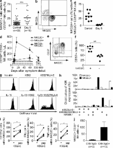
          Acute hantavirus infection in humans triggers a rapid expansion and long-term persistence of NK cells.

          Abstract

          Natural killer (NK) cells are known to mount a rapid response to several virus infections. In experimental models of acute viral infection, this response has been characterized by prompt NK cell activation and expansion followed by rapid contraction. In contrast to experimental model systems, much less is known about NK cell responses to acute viral infections in humans. We demonstrate that NK cells can rapidly expand and persist at highly elevated levels for >60 d after human hantavirus infection. A large part of the expanding NK cells expressed the activating receptor NKG2C and were functional in terms of expressing a licensing inhibitory killer cell immunoglobulin-like receptor (KIR) and ability to respond to target cell stimulation. These results demonstrate that NK cells can expand and remain elevated in numbers for a prolonged period of time in humans after a virus infection. In time, this response extends far beyond what is considered normal for an innate immune response.

          Related collections

          Most cited references43

          • Record: found
          • Abstract: found
          • Article: not found

          HLA-E binds to natural killer cell receptors CD94/NKG2A, B and C.

          The protein HLA-E is a non-classical major histocompatibility complex (MHC) molecule of limited sequence variability. Its expression on the cell surface is regulated by the binding of peptides derived from the signal sequence of some other MHC class I molecules. Here we report the identification of ligands for HLA-E. We constructed tetramers in which recombinant HLA-E and beta2-microglobulin were refolded with an MHC leader-sequence peptide, biotinylated, and conjugated to phycoerythrin-labelled Extravidin. This HLA-E tetramer bound to natural killer (NK) cells and a small subset of T cells from peripheral blood. On transfectants, the tetramer bound to the CD94/NKG2A, CD94/NKGK2B and CD94/NKG2C NK cell receptors, but did not bind to the immunoglobulin family of NK cell receptors (KIR). Surface expression of HLA-E was enough to protect target cells from lysis by CD94/NKG2A+ NK-cell clones. A subset of HLA class I alleles has been shown to inhibit killing by CD94/NKG2A+ NK-cell clones. Only the HLA alleles that possess a leader peptide capable of upregulating HLA-E surface expression confer resistance to NK-cell-mediated lysis, implying that their action is mediated by HLA-E, the predominant ligand for the NK cell inhibitory receptor CD94/NKG2A.
            Bookmark
            • Record: found
            • Abstract: found
            • Article: not found

            Immunology of hepatitis B virus and hepatitis C virus infection.

            More than 500 million people worldwide are persistently infected with the hepatitis B virus (HBV) and/or hepatitis C virus (HCV) and are at risk of developing chronic liver disease, cirrhosis and hepatocellular carcinoma. Despite many common features in the pathogenesis of HBV- and HCV-related liver disease, these viruses markedly differ in their virological properties and in their immune escape and survival strategies. This review assesses recent advances in our understanding of viral hepatitis, contrasts mechanisms of virus-host interaction in acute hepatitis B and hepatitis C, and outlines areas for future studies.
              Bookmark
              • Record: found
              • Abstract: found
              • Article: not found

              Expression patterns of NKG2A, KIR, and CD57 define a process of CD56dim NK-cell differentiation uncoupled from NK-cell education.

              Natural killer (NK) cells are lymphocytes of the innate immune system that, following differentiation from CD56(bright) to CD56(dim) cells, have been thought to retain fixed functional and phenotypic properties throughout their lifespan. In contrast to this notion, we here show that CD56(dim) NK cells continue to differentiate. During this process, they lose expression of NKG2A, sequentially acquire inhibitory killer cell inhibitory immunoglobulin-like receptors and CD57, change their expression patterns of homing molecules, and display a gradual decline in proliferative capacity. All cellular intermediates of this process are represented in varying proportions at steady state and appear, over time, during the reconstitution of the immune system, as demonstrated in humanized mice and in patients undergoing hematopoietic stem cell transplantation. CD56(dim) NK-cell differentiation, and the associated functional imprint, occurs independently of NK-cell education by interactions with self-human leukocyte antigen class I ligands and is an essential part of the formation of human NK-cell repertoires.
                Bookmark

                Author and article information

                Journal
                J Exp Med
                J. Exp. Med
                jem
                The Journal of Experimental Medicine
                The Rockefeller University Press
                0022-1007
                1540-9538
                17 January 2011
                : 208
                : 1
                : 13-21
                Affiliations
                [1 ]Center for Infectious Medicine, Department of Medicine, Karolinska Institutet, Karolinska University Hospital Huddinge, 141 86 Stockholm, Sweden
                [2 ]Department of Clinical Microbiology, Division of Infectious Diseases , and [3 ]Department of Clinical Microbiology, Division of Virology, Umeå University, 901 85 Umeå, Sweden
                [4 ]Department of Microbiology, Tumor and Cell Biology, Karolinska Institutet, 171 77 Stockholm, Sweden
                [5 ]Center for Microbiological Preparedness, Swedish Institute for Infectious Disease Control, 171 82 Solna, Sweden
                [6 ]Centre d’Immunologie de Marseille-Luminy, Centre National de la Recherche Scientifique–Institut National de la Santé et de la Recherche Médicale, Université de la Méditerranée Campus de Luminy, 13288 Marseille Cedex 09, France
                Author notes
                CORRESPONDENCE Hans-Gustaf Ljunggren: hans-gustaf.ljunggren@ 123456ki.se
                Article
                20100762
                10.1084/jem.20100762
                3023129
                21173105
                e2441882-9c20-4b23-9b5d-fcf8ade0ae01
                © 2011 Björkström et al.

                This article is distributed under the terms of an Attribution–Noncommercial–Share Alike–No Mirror Sites license for the first six months after the publication date (see http://www.rupress.org/terms). After six months it is available under a Creative Commons License (Attribution–Noncommercial–Share Alike 3.0 Unported license, as described at http://creativecommons.org/licenses/by-nc-sa/3.0/).

                History
                : 19 April 2010
                : 12 November 2010
                Categories
                Brief Definitive Report

                Medicine
                Medicine

                Comments

                Comment on this article