857
views
0
recommends
+1 Recommend
0 collections
    0
    shares
      • Record: found
      • Abstract: found
      • Article: found
      Is Open Access

      Rapid Healing of Cutaneous Leishmaniasis by High-Frequency Electrocauterization and Hydrogel Wound Care with or without DAC N-055: A Randomized Controlled Phase IIa Trial in Kabul

      research-article

      Read this article at

      Bookmark
          There is no author summary for this article yet. Authors can add summaries to their articles on ScienceOpen to make them more accessible to a non-specialist audience.

          Abstract

          Background

          Anthroponotic cutaneous leishmaniasis (CL) due to Leishmania (L.) tropica infection is a chronic, frequently disfiguring skin disease with limited therapeutic options. In endemic countries healing of ulcerative lesions is often delayed by bacterial and/or fungal infections. Here, we studied a novel therapeutic concept to prevent superinfections, accelerate wound closure, and improve the cosmetic outcome of ACL.

          Methodology/Principal Findings

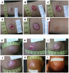
          From 2004 to 2008 we performed a two-armed, randomized, double-blinded, phase IIa trial in Kabul, Afghanistan, with patients suffering from L. tropica CL. The skin lesions were treated with bipolar high-frequency electrocauterization (EC) followed by daily moist-wound-treatment (MWT) with polyacrylate hydrogel with (group I) or without (group II) pharmaceutical sodium chlorite (DAC N-055). Patients below age 5, with facial lesions, pregnancy, or serious comorbidities were excluded. The primary, photodocumented outcome was the time needed for complete lesion epithelialization. Biopsies for parasitological and (immuno)histopathological analyses were taken prior to EC (1 st), after wound closure (2 nd) and after 6 months (3 rd). The mean duration for complete wound closure was short and indifferent in group I (59 patients, 43.1 d) and II (54 patients, 42 d; p = 0.83). In patients with Leishmania-positive 2 nd biopsies DAC N-055 caused a more rapid wound epithelialization (37.2 d vs. 58.3 d; p = 0.08). Superinfections occurred in both groups at the same rate (8.8%). Except for one patient, reulcerations (10.2% in group I, 18.5% in group II; p = 0.158) were confined to cases with persistent high parasite loads after healing. In vitro, DAC N-055 showed a leishmanicidal effect on pro- and amastigotes.

          Conclusions/Significance

          Compared to previous results with intralesional antimony injections, the EC plus MWT protocol led to more rapid wound closure. The tentatively lower rate of relapses and the acceleration of wound closure in a subgroup of patients with parasite persistence warrant future studies on the activity of DAC N-055.

          Trial Registration

          ClinicalTrails.gov NCT00947362

          Author Summary

          In many countries of the Middle East such as Afghanistan, cutaneous leishmaniasis is a highly prevalent, chronic and stigmatizing skin disease. Poor hygiene conditions frequently aggravate the lesions due to bacterial and fungal superinfections. Classical treatments with injections of pentavalent antimony are hampered by costs, side effects, resistance development, supply and manufactural quality problems. In the present study on Afghan patients with Leishmania tropica-induced skin lesions we evaluated the clinical effect of an initial removal of lesion tissue by electrocoagulation using a bipolar high-frequency electrosurgery instrument, followed by daily moist wound treatment with or without a preparation of pharmaceutical sodium chlorite (DAC N-055). DAC N-055 is a compound with anti-infective, immunomodulatory and tissue repair-promoting effects. Our analysis revealed that the carefully performed moist wound treatment led to a rapid healing of the wounds within an average period of 6 weeks, even in the absence of the sodium chlorite preparation. This is considerably faster than the time spans previously reported for local or systemic antimony treatment. We believe that the current standard for local care of chronic wounds should also be applied to Leishmania skin lesions. If combined with an initial single high-frequency electrocoagulation, it is a highly effective, inexpensive and well-tolerated treatment option for cutaneous leishmaniasis.

          Related collections

          Most cited references71

          • Record: found
          • Abstract: found
          • Article: not found

          Regulation of wound healing by growth factors and cytokines.

          Cutaneous wound healing is a complex process involving blood clotting, inflammation, new tissue formation, and finally tissue remodeling. It is well described at the histological level, but the genes that regulate skin repair have only partially been identified. Many experimental and clinical studies have demonstrated varied, but in most cases beneficial, effects of exogenous growth factors on the healing process. However, the roles played by endogenous growth factors have remained largely unclear. Initial approaches at addressing this question focused on the expression analysis of various growth factors, cytokines, and their receptors in different wound models, with first functional data being obtained by applying neutralizing antibodies to wounds. During the past few years, the availability of genetically modified mice has allowed elucidation of the function of various genes in the healing process, and these studies have shed light onto the role of growth factors, cytokines, and their downstream effectors in wound repair. This review summarizes the results of expression studies that have been performed in rodents, pigs, and humans to localize growth factors and their receptors in skin wounds. Most importantly, we also report on genetic studies addressing the functions of endogenous growth factors in the wound repair process.
            Bookmark
            • Record: found
            • Abstract: not found
            • Article: not found

            Formation of the scab and the rate of epithelization of superficial wounds in the skin of the young domestic pig.

              Bookmark
              • Record: found
              • Abstract: found
              • Article: not found

              Electrical signals control wound healing through phosphatidylinositol-3-OH kinase-gamma and PTEN.

              Wound healing is essential for maintaining the integrity of multicellular organisms. In every species studied, disruption of an epithelial layer instantaneously generates endogenous electric fields, which have been proposed to be important in wound healing. The identity of signalling pathways that guide both cell migration to electric cues and electric-field-induced wound healing have not been elucidated at a genetic level. Here we show that electric fields, of a strength equal to those detected endogenously, direct cell migration during wound healing as a prime directional cue. Manipulation of endogenous wound electric fields affects wound healing in vivo. Electric stimulation triggers activation of Src and inositol-phospholipid signalling, which polarizes in the direction of cell migration. Notably, genetic disruption of phosphatidylinositol-3-OH kinase-gamma (PI(3)Kgamma) decreases electric-field-induced signalling and abolishes directed movements of healing epithelium in response to electric signals. Deletion of the tumour suppressor phosphatase and tensin homolog (PTEN) enhances signalling and electrotactic responses. These data identify genes essential for electrical-signal-induced wound healing and show that PI(3)Kgamma and PTEN control electrotaxis.
                Bookmark

                Author and article information

                Contributors
                Role: Editor
                Journal
                PLoS Negl Trop Dis
                PLoS Negl Trop Dis
                plos
                plosntds
                PLoS Neglected Tropical Diseases
                Public Library of Science (San Francisco, USA )
                1935-2727
                1935-2735
                February 2014
                13 February 2014
                : 8
                : 2
                : e2694
                Affiliations
                [1 ]Mikrobiologisches Institut – Klinische Mikrobiologie, Immunologie und Hygiene, Friedrich-Alexander-Universität (FAU) Erlangen-Nürnberg and Universitätsklinikum Erlangen, Erlangen, Germany
                [2 ]Leishmania Clinic, German Medical Service (NGO), Darwaze-e-Lahory, Kabul, Afghanistan
                [3 ]Waisenmedizin e.V. Promoting Access to Care with Essential Medicine, Non-Profit Organization, Freiburg, Germany
                National Institutes of Health, United States of America
                Author notes

                The authors have declared that no competing interests exist.

                Conceived and designed the experiments: KWS RS FMA US CB. Performed the experiments: AFJ US RS PW FM KWS. Analyzed the data: AFJ US PW HCS CB KWS. Contributed reagents/materials/analysis tools: KWS US CB FMA. Wrote the paper: CB US AFJ KWS. Final approval of the manuscript version to be published: AFJ US RS PW FM HCS FMA CB KWS.

                [¤a]

                Current address: Department of Cardiovascular and Thoracic Surgery, Universitätsklinikum Göttingen and Georg-August-Universität, Göttingen, Germany.

                [¤b]

                Current address: Christusträger Communität, Gut Rallingen am Thunersee, Merligen, Switzerland.

                [¤c]

                Current address: German Medical Diagnostic Center Kabul DK-GMDC Ltd., Kabul, Afghanistan.

                ¶ These authors are joint senior authors on this work.

                ‡ Meanwhile retired.

                Article
                PNTD-D-13-01363
                10.1371/journal.pntd.0002694
                3923720
                c0f5dbd7-42b6-4357-9970-b7e774961dc0
                Copyright @ 2014

                This is an open-access article distributed under the terms of the Creative Commons Attribution License, which permits unrestricted use, distribution, and reproduction in any medium, provided the original author and source are credited.

                History
                : 8 September 2013
                : 29 December 2013
                Page count
                Pages: 13
                Funding
                This study was supported by the German-Academic Exchange Service (travel grants to KWS, AFJ and FM), the Senior Expert Service (travel grants to KWS) and the Interdisciplinary Center of Clinical Research at the Universitätsklinikum Erlangen (IZKF, project A49 to US and CB). The funders had no role in study design, data collection and analysis, decision to publish, or preparation of the manuscript.
                Categories
                Research Article
                Medicine
                Infectious diseases
                Neglected tropical diseases
                Leishmaniasis
                Skin infections

                Infectious disease & Microbiology
                Infectious disease & Microbiology

                Comments

                Comment on this article