17
views
0
recommends
+1 Recommend
0 collections
    0
    shares
      • Record: found
      • Abstract: found
      • Article: found
      Is Open Access

      ATP synthase: Evolution, energetics, and membrane interactions

      review-article

      Read this article at

      Bookmark
          There is no author summary for this article yet. Authors can add summaries to their articles on ScienceOpen to make them more accessible to a non-specialist audience.

          Abstract

          This review outlines a holistic framework for studying ATP synthase and emphasizes the importance of considering interactions with the lipid environment in shaping the function and evolutionary history of membrane proteins.

          Abstract

          The synthesis of ATP, life’s “universal energy currency,” is the most prevalent chemical reaction in biological systems and is responsible for fueling nearly all cellular processes, from nerve impulse propagation to DNA synthesis. ATP synthases, the family of enzymes that carry out this endless task, are nearly as ubiquitous as the energy-laden molecule they are responsible for making. The F-type ATP synthase (F-ATPase) is found in every domain of life and has facilitated the survival of organisms in a wide range of habitats, ranging from the deep-sea thermal vents to the human intestine. Accordingly, there has been a large amount of work dedicated toward understanding the structural and functional details of ATP synthases in a wide range of species. Less attention, however, has been paid toward integrating these advances in ATP synthase molecular biology within the context of its evolutionary history. In this review, we present an overview of several structural and functional features of the F-type ATPases that vary across taxa and are purported to be adaptive or otherwise evolutionarily significant: ion channel selectivity, rotor ring size and stoichiometry, ATPase dimeric structure and localization in the mitochondrial inner membrane, and interactions with membrane lipids. We emphasize the importance of studying these features within the context of the enzyme’s particular lipid environment. Just as the interactions between an organism and its physical environment shape its evolutionary trajectory, ATPases are impacted by the membranes within which they reside. We argue that a comprehensive understanding of the structure, function, and evolution of membrane proteins—including ATP synthase—requires such an integrative approach.

          Related collections

          Most cited references234

          • Record: found
          • Abstract: found
          • Article: not found

          Membrane lipids: where they are and how they behave.

          Throughout the biological world, a 30 A hydrophobic film typically delimits the environments that serve as the margin between life and death for individual cells. Biochemical and biophysical findings have provided a detailed model of the composition and structure of membranes, which includes levels of dynamic organization both across the lipid bilayer (lipid asymmetry) and in the lateral dimension (lipid domains) of membranes. How do cells apply anabolic and catabolic enzymes, translocases and transporters, plus the intrinsic physical phase behaviour of lipids and their interactions with membrane proteins, to create the unique compositions and multiple functionalities of their individual membranes?
            Bookmark
            • Record: found
            • Abstract: found
            • Article: found
            Is Open Access

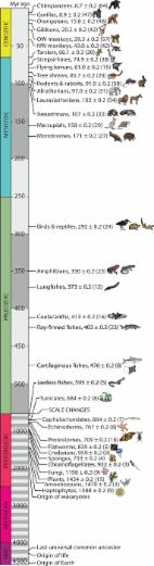
            Tree of Life Reveals Clock-Like Speciation and Diversification

            Genomic data are rapidly resolving the tree of living species calibrated to time, the timetree of life, which will provide a framework for research in diverse fields of science. Previous analyses of taxonomically restricted timetrees have found a decline in the rate of diversification in many groups of organisms, often attributed to ecological interactions among species. Here, we have synthesized a global timetree of life from 2,274 studies representing 50,632 species and examined the pattern and rate of diversification as well as the timing of speciation. We found that species diversity has been mostly expanding overall and in many smaller groups of species, and that the rate of diversification in eukaryotes has been mostly constant. We also identified, and avoided, potential biases that may have influenced previous analyses of diversification including low levels of taxon sampling, small clade size, and the inclusion of stem branches in clade analyses. We found consistency in time-to-speciation among plants and animals, ∼2 My, as measured by intervals of crown and stem species times. Together, this clock-like change at different levels suggests that speciation and diversification are processes dominated by random events and that adaptive change is largely a separate process.
              Bookmark
              • Record: found
              • Abstract: found
              • Article: not found

              Crystal structure of a bacterial homologue of Na+/Cl--dependent neurotransmitter transporters.

              Na+/Cl--dependent transporters terminate synaptic transmission by using electrochemical gradients to drive the uptake of neurotransmitters, including the biogenic amines, from the synapse to the cytoplasm of neurons and glia. These transporters are the targets of therapeutic and illicit compounds, and their dysfunction has been implicated in multiple diseases of the nervous system. Here we present the crystal structure of a bacterial homologue of these transporters from Aquifex aeolicus, in complex with its substrate, leucine, and two sodium ions. The protein core consists of the first ten of twelve transmembrane segments, with segments 1-5 related to 6-10 by a pseudo-two-fold axis in the membrane plane. Leucine and the sodium ions are bound within the protein core, halfway across the membrane bilayer, in an occluded site devoid of water. The leucine and ion binding sites are defined by partially unwound transmembrane helices, with main-chain atoms and helix dipoles having key roles in substrate and ion binding. The structure reveals the architecture of this important class of transporter, illuminates the determinants of substrate binding and ion selectivity, and defines the external and internal gates.
                Bookmark

                Author and article information

                Journal
                J Gen Physiol
                J Gen Physiol
                jgp
                The Journal of General Physiology
                Rockefeller University Press
                0022-1295
                1540-7748
                02 November 2020
                23 September 2020
                : 152
                : 11
                : e201912475
                Affiliations
                [1 ]Center for Studies in Physics and Biology, The Rockefeller University, New York, NY
                [2 ]All Souls College, University of Oxford, Oxford, UK
                [3 ]Department of Chemistry and Biochemistry, University of California San Diego, La Jolla, CA
                [4 ]Department of Mechanical and Aerospace Engineering, University of California San Diego, La Jolla, CA
                Author notes
                Correspondence to Jasmine A. Nirody: jnirody@ 123456rockefeller.edu
                Padmini Rangamani: prangamani@ 123456ucsd.edu
                Author information
                https://orcid.org/0000-0002-0461-1031
                https://orcid.org/0000-0001-9706-4294
                https://orcid.org/0000-0001-5953-4347
                Article
                jgp.201912475
                10.1085/jgp.201912475
                7594442
                32966553
                bca12323-5393-4105-aef3-09e4fa9b83e7
                © 2020 Nirody et al.

                This article is distributed under the terms of an Attribution–Noncommercial–Share Alike–No Mirror Sites license for the first six months after the publication date (see http://www.rupress.org/terms/). After six months it is available under a Creative Commons License (Attribution–Noncommercial–Share Alike 4.0 International license, as described at https://creativecommons.org/licenses/by-nc-sa/4.0/).

                History
                : 16 June 2020
                : 24 August 2020
                Page count
                Pages: 18
                Funding
                Funded by: James S. McDonnell Foundation, DOI http://dx.doi.org/10.13039/100000913;
                Funded by: The Rockefeller University, DOI http://dx.doi.org/10.13039/100012007;
                Funded by: All Souls College, DOI http://dx.doi.org/10.13039/501100000524;
                Funded by: National Science Foundation, DOI http://dx.doi.org/10.13039/100000001;
                Award ID: MCB-1715681
                Funded by: Air Force Office of Scientific Research, DOI http://dx.doi.org/10.13039/100000181;
                Categories
                Review
                Protein Structure and Dynamics
                Lipids and Membranes
                Biophysics

                Anatomy & Physiology
                Anatomy & Physiology

                Comments

                Comment on this article