8
views
0
recommends
+1 Recommend
0 collections
    0
    shares
      • Record: found
      • Abstract: found
      • Article: found
      Is Open Access

      Micromechanical modeling of the contact stiffness of an osseointegrated bone–implant interface

      research-article

      Read this article at

      Bookmark
          There is no author summary for this article yet. Authors can add summaries to their articles on ScienceOpen to make them more accessible to a non-specialist audience.

          Abstract

          Background

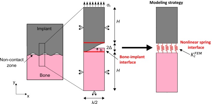
          The surgical success of cementless implants is determined by the evolution of the biomechanical properties of the bone–implant interface (BII). One difficulty to model the biomechanical behavior of the BII comes from the implant surface roughness and from the partial contact between bone tissue and the implant. The determination of the constitutive law of the BII would be of interest in the context of implant finite element (FE) modeling to take into account the imperfect characteristics of the BII. The aim of the present study is to determine an effective contact stiffness \documentclass[12pt]{minimal} \usepackage{amsmath} \usepackage{wasysym} \usepackage{amsfonts} \usepackage{amssymb} \usepackage{amsbsy} \usepackage{mathrsfs} \usepackage{upgreek} \setlength{\oddsidemargin}{-69pt} \begin{document}$$\left( {K_{c}^{\text{FEM}} } \right)$$\end{document} of an osseointegrated BII accounting for its micromechanical features such as surface roughness, bone–implant contact ratio (BIC) and periprosthetic bone properties. To do so, a 2D FE model of the BII under normal contact conditions was developed and was used to determine the behavior of \documentclass[12pt]{minimal} \usepackage{amsmath} \usepackage{wasysym} \usepackage{amsfonts} \usepackage{amssymb} \usepackage{amsbsy} \usepackage{mathrsfs} \usepackage{upgreek} \setlength{\oddsidemargin}{-69pt} \begin{document}$$K_{c}^{\text{FEM}}$$\end{document} .

          Results

          The model is validated by comparison with three analytical schemes based on micromechanical homogenization including two Lekesiz’s models (considering interacting and non-interacting micro-cracks) and a Kachanov’s model. \documentclass[12pt]{minimal} \usepackage{amsmath} \usepackage{wasysym} \usepackage{amsfonts} \usepackage{amssymb} \usepackage{amsbsy} \usepackage{mathrsfs} \usepackage{upgreek} \setlength{\oddsidemargin}{-69pt} \begin{document}$$K_{c}^{\text{FEM}}$$\end{document} is found to be comprised between 10 13 and 10 15 N/m 3 according to the properties of the BII. \documentclass[12pt]{minimal} \usepackage{amsmath} \usepackage{wasysym} \usepackage{amsfonts} \usepackage{amssymb} \usepackage{amsbsy} \usepackage{mathrsfs} \usepackage{upgreek} \setlength{\oddsidemargin}{-69pt} \begin{document}$$K_{c}^{\text{FEM}}$$\end{document} is shown to increase nonlinearly as a function of the BIC and to decrease as a function of the roughness amplitude for high BIC values (above around 20%). Moreover, \documentclass[12pt]{minimal} \usepackage{amsmath} \usepackage{wasysym} \usepackage{amsfonts} \usepackage{amssymb} \usepackage{amsbsy} \usepackage{mathrsfs} \usepackage{upgreek} \setlength{\oddsidemargin}{-69pt} \begin{document}$$K_{c}^{\text{FEM}}$$\end{document} decreases as a function of the roughness wavelength and increases linearly as a function of the Young’s modulus of periprosthetic bone tissue.

          Conclusions

          These results open new paths in implant biomechanical modeling since this model may be used in future macroscopic finite element models modeling the bone–implant system to replace perfectly rigid BII conditions.

          Related collections

          Most cited references52

          • Record: found
          • Abstract: found
          • Article: not found

          Osteoinduction, osteoconduction and osseointegration.

          Osteoinduction is the process by which osteogenesis is induced. It is a phenomenon regularly seen in any type of bone healing process. Osteoinduction implies the recruitment of immature cells and the stimulation of these cells to develop into preosteoblasts. In a bone healing situation such as a fracture, the majority of bone healing is dependent on osteoinduction. Osteoconduction means that bone grows on a surface. This phenomenon is regularly seen in the case of bone implants. Implant materials of low biocompatibility such as copper, silver and bone cement shows little or no osteoconduction. Osseointegration is the stable anchorage of an implant achieved by direct bone-to-implant contact. In craniofacial implantology, this mode of anchorage is the only one for which high success rates have been reported. Osseointegration is possible in other parts of the body, but its importance for the anchorage of major arthroplasties is under debate. Ingrowth of bone in a porous-coated prosthesis may or may not represent osseointegration.
            Bookmark
            • Record: found
            • Abstract: found
            • Article: not found

            Observations on the effect of movement on bone ingrowth into porous-surfaced implants.

            Although porous-surfaced orthopedic implants have been designed for fixation by bone ingrowth, there is clinical evidence that this does not always occur. Initial implant movement relative to host bone can result in attachment by a nonmineralized fibrous connective tissue layer. The ranges of movement that result in either bone or fibrous connective tissue fixation are observed in dogs in two independent studies. Experimentally, bone ingrowth can occur in the presence of some movement, albeit very small (up to 28 mu), while excess movement (150 mu or more) can result in attachment by mature connective tissue ingrowth.
              Bookmark
              • Record: found
              • Abstract: found
              • Article: not found

              Early bone formation adjacent to rough and turned endosseous implant surfaces. An experimental study in the dog.

              To validate a proposed model (Berglundh et al. 2003) and to evaluate the rate and degree of osseointegration at turned (T) and sand blasted and acid etched (SLA) implant surfaces during early phases of healing. The devices used for the study of early healing had a geometry that corresponded to that of a solid screw implant with either a SLA or a T surface configuration. A circumferential trough had been prepared within the thread region (intra-osseous portion) that established a geometrically well-defined wound chamber. Twenty Labrador dogs received totally 160 experimental devices to allow the evaluation of healing between 2 h and 12 weeks. Both ground and decalcified sections were prepared from mesial/distal and buccal/lingual device sites. Histometric and morphometric analyses of the ground sections and morphometric analysis of the tissue components in decalcified sections were performed. The ground sections provided an overview of the various phases of tissue formation, while the decalcified, thin sections enabled a more detailed study of events involved in bone tissue modeling and remodeling for both SLA and T surfaces. The initially empty wound chamber became occupied with a coagulum and a granulation tissue that was replaced by a provisional matrix. The process of bone formation started already during the first week. The newly formed bone present at the lateral border of the cut bony bed appeared to be continuous with the parent bone, but on the SLA surface woven bone was also found at a distance from the parent bone. Parallel-fibered and/or lamellar bone as well as bone marrow replaced this primary bone after 4 weeks. In the SLA chambers, more bone-to-device contact, more initial woven bone and earlier lamellar bone formation was found than in the T chambers. Osseointegration represents a dynamic process both during its establishment and its maintenance. While healing showed similar characteristics with resorptive and appositional events for both SLA and T surfaces, the rate and degree of osseointegration were superior for the SLA compared with the T chambers.
                Bookmark

                Author and article information

                Contributors
                mariraffa@hotmail.com
                vu-hieu.nguyen@u-pec.fr
                guillaume.haiat@cnrs.fr
                Journal
                Biomed Eng Online
                Biomed Eng Online
                BioMedical Engineering OnLine
                BioMed Central (London )
                1475-925X
                3 December 2019
                3 December 2019
                2019
                : 18
                : 114
                Affiliations
                GRID grid.462588.5, CNRS, Laboratoire Modélisation et Simulation Multi Echelle, , MSME, UMR CNRS 8208, ; 61 Avenue du Général de Gaulle, 94010 Créteil, France
                Author information
                http://orcid.org/0000-0002-4062-7153
                http://orcid.org/0000-0003-1724-9083
                Article
                733
                10.1186/s12938-019-0733-3
                6889538
                31796076
                bb26abb1-3322-4fc7-a86c-cd0b77e0914b
                © The Author(s) 2019

                Open AccessThis article is distributed under the terms of the Creative Commons Attribution 4.0 International License ( http://creativecommons.org/licenses/by/4.0/), which permits unrestricted use, distribution, and reproduction in any medium, provided you give appropriate credit to the original author(s) and the source, provide a link to the Creative Commons license, and indicate if changes were made. The Creative Commons Public Domain Dedication waiver (http://creativecommons.org/publicdomain/zero/1.0/) applies to the data made available in this article, unless otherwise stated.

                History
                : 3 June 2019
                : 21 November 2019
                Funding
                Funded by: European research council
                Award ID: 682001
                Award Recipient :
                Categories
                Research
                Custom metadata
                © The Author(s) 2019

                Biomedical engineering
                bone–implant interface,contact,roughness,homogenization,finite element modeling

                Comments

                Comment on this article