9
views
0
recommends
+1 Recommend
0 collections
    0
    shares
      • Record: found
      • Abstract: found
      • Article: not found

      Expression of HDAC2 but Not HDAC1 Transcript Is Reduced in Dorsolateral Prefrontal Cortex of Patients with Schizophrenia

      research-article

      Read this article at

      ScienceOpenPublisherPMC
      Bookmark
          There is no author summary for this article yet. Authors can add summaries to their articles on ScienceOpen to make them more accessible to a non-specialist audience.

          Abstract

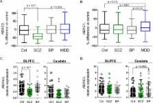
          Postmortem brain studies support dysregulated expression of the histone deacetylase enzymes, HDAC1 and HDAC2, as a central feature in diseases including schizophrenia, bipolar disorder, and depression. Our objective was to investigate HDAC expression in a large postmortem sample set representing healthy and disease brains. We used >700 well-characterized samples from patients diagnosed with schizophrenia ( n = 175), major depressive disorder ( n = 135), and bipolar disorder ( n = 61) to measure HDAC1 and HDAC2 transcript levels by quantitative real-time PCR in dorsolateral prefrontal cortex (DLPFC) and caudate compared to control samples. HDAC expression was calculated relative to the geometric mean of β-2-microglobulin, β-glucuronidase, and β-actin. In adult-age DLPFC, HDAC2 was decreased by 34% in schizophrenia samples compared to controls ( p < 10 –4). HDAC2 was significantly upregulated in major depressive disorder samples by 17% versus controls ( p = 0.002). Neither smoking history nor therapeutic drugs impacted HDAC2 levels and no HDAC1 patient-control differences were observed. In caudate, HDAC levels were unchanged between patient and control groups. In control DLPFC, age fetal week 14 to 97 years ( n = 326), both HDAC1 and HDAC2 levels sharply declined around birth and stabilized thereafter. Using by far the largest postmortem sample set on this topic, our major finding (decreased HDAC2 transcript) showed notable specificity in disease (schizophrenia but not major depressive disorder), HDAC subtype ( HDAC2 but not HDAC1) and brain region (DLPFC but not caudate). These differences shape understanding of regional components of neural circuitry in the diseased brain and set a benchmark to quantify HDAC density and distribution using in vivo neuroimaging tools.

          Related collections

          Most cited references29

          • Record: found
          • Abstract: found
          • Article: not found

          Temporal dynamics and genetic control of transcription in the human prefrontal cortex.

          Previous investigations have combined transcriptional and genetic analyses in human cell lines, but few have applied these techniques to human neural tissue. To gain a global molecular perspective on the role of the human genome in cortical development, function and ageing, we explore the temporal dynamics and genetic control of transcription in human prefrontal cortex in an extensive series of post-mortem brains from fetal development through ageing. We discover a wave of gene expression changes occurring during fetal development which are reversed in early postnatal life. One half-century later in life, this pattern of reversals is mirrored in ageing and in neurodegeneration. Although we identify thousands of robust associations of individual genetic polymorphisms with gene expression, we also demonstrate that there is no association between the total extent of genetic differences between subjects and the global similarity of their transcriptional profiles. Hence, the human genome produces a consistent molecular architecture in the prefrontal cortex, despite millions of genetic differences across individuals and races. To enable further discovery, this entire data set is freely available (from Gene Expression Omnibus: accession GSE30272; and dbGaP: accession phs000417.v1.p1) and can also be interrogated via a biologist-friendly stand-alone application (http://www.libd.org/braincloud).
            Bookmark
            • Record: found
            • Abstract: found
            • Article: not found

            Antidepressant actions of histone deacetylase inhibitors.

            Persistent symptoms of depression suggest the involvement of stable molecular adaptations in brain, which may be reflected at the level of chromatin remodeling. We find that chronic social defeat stress in mice causes a transient decrease, followed by a persistent increase, in levels of acetylated histone H3 in the nucleus accumbens, an important limbic brain region. This persistent increase in H3 acetylation is associated with decreased levels of histone deacetylase 2 (HDAC2) in the nucleus accumbens. Similar effects were observed in the nucleus accumbens of depressed humans studied postmortem. These changes in H3 acetylation and HDAC2 expression mediate long-lasting positive neuronal adaptations, since infusion of HDAC inhibitors into the nucleus accumbens, which increases histone acetylation, exerts robust antidepressant-like effects in the social defeat paradigm and other behavioral assays. HDAC inhibitor [N-(2-aminophenyl)-4-[N-(pyridine-3-ylmethoxy-carbonyl)aminomethyl]benzamide (MS-275)] infusion also reverses the effects of chronic defeat stress on global patterns of gene expression in the nucleus accumbens, as determined by microarray analysis, with striking similarities to the effects of the standard antidepressant fluoxetine. Stress-regulated genes whose expression is normalized selectively by MS-275 may provide promising targets for the future development of novel antidepressant treatments. Together, these findings provide new insight into the underlying molecular mechanisms of depression and antidepressant action, and support the antidepressant potential of HDAC inhibitors and perhaps other agents that act at the level of chromatin structure.
              Bookmark
              • Record: found
              • Abstract: found
              • Article: not found

              Regulation of the GABA cell phenotype in hippocampus of schizophrenics and bipolars.

              GABAergic dysfunction is present in the hippocampus in schizophrenia (SZ) and bipolar disorder (BD). The trisynaptic pathway was "deconstructed" into various layers of sectors CA3/2 and CA1 and gene expression profiling performed. Network association analysis was used to uncover genes that may be related to regulation of glutamate decarboxylase 67 (GAD(67)), a marker for this system that has been found by many studies to show decreased expression in SZs and BDs. The most striking change was a down-regulation of GAD(67) in the stratum oriens (SO) of CA2/3 in both groups; CA1 only showed changes in the SO of schizophrenics. The network generated for GAD(67) contained 25 genes involved in the regulation of kainate receptors, TGF-beta and Wnt signaling, as well as transcription factors involved in cell growth and differentiation. In SZs, IL-1beta, (GRIK2/3), TGF-beta2, TGF-betaR1, histone deacetylase 1 (HDAC1), death associated protein (DAXX), and cyclin D2 (CCND2) were all significantly up-regulated, whereas in BDs, PAX5, Runx2, LEF1, TLE1, and CCND2 were significantly down-regulated. In the SO of CA1 of BDs, where GAD67 showed no expression change, TGF-beta and Wnt signaling genes were all up-regulated, but other transcription factors showed no change in expression. In other layers/sectors, BDs showed no expression changes in these GAD(67) network genes. Overall, these results are consistent with the hypothesis that decreased expression of GAD(67) may be associated with an epigenetic mechanism in SZ. In BD, however, a suppression of transcription factors involved in cell differentiation may contribute to GABA dysfunction.
                Bookmark

                Author and article information

                Journal
                ACS Chem Neurosci
                ACS Chem Neurosci
                cn
                acncdm
                ACS Chemical Neuroscience
                American Chemical Society
                1948-7193
                28 November 2016
                15 March 2017
                : 8
                : 3
                : 662-668
                Affiliations
                []Athinoula A. Martinos Center, Department of Radiology, Massachusetts General Hospital, Harvard Medical School , Charlestown, Massachusetts 02129, United States
                []Human Brain Collection Core, Division of Intramural Research Programs, National Institute of Mental Health, National Institutes of Health, Bethesda, Maryland 20892, United States
                [§ ]National Institute on Alcohol Abuse and Alcoholism, National Institutes of Health, Bethesda, Maryland 20892, United States
                []Molecular Imaging Branch, National Institute of Mental Health, National Institutes of Health, Bethesda, Maryland 20892, United States
                Author notes
                [* ]Mailing address: Athinoula A. Martinos Center for Biomedical Imaging, Harvard Medical School, Massachusetts General Hospital, Building 149, 13th Street, Suite 2301 Charlestown, MA 02129, USA. E-mail: fschroeder@ 123456mgh.harvard.edu . Telephone: 1-617-643-7811.
                Article
                10.1021/acschemneuro.6b00372
                5436730
                27959513
                b0b39018-3c18-4b8f-9fbd-cb93c1163547
                Copyright © 2016 American Chemical Society

                This is an open access article published under an ACS AuthorChoice License, which permits copying and redistribution of the article or any adaptations for non-commercial purposes.

                History
                : 01 November 2016
                : 28 November 2016
                Categories
                Research Article
                Custom metadata
                cn6b00372
                cn-2016-00372k

                Neurosciences
                epigenetics,chromatin,schizophrenia,mood disorders unipolar,mood disorders bipolar,neuropsychiatry

                Comments

                Comment on this article