33
views
0
recommends
+1 Recommend
2 collections
    0
    shares
      • Record: found
      • Abstract: found
      • Article: found
      Is Open Access

      Persistence and decay of human antibody responses to the receptor binding domain of SARS-CoV-2 spike protein in COVID-19 patients

      research-article
      a , b , c , a , a , a , a , a , a , a , d , d , d , d , a , e , e , d , a , b , f , c , g , g , e , e , e , d , a , b , d , a , b , a , e , g , e , f , c , e , a , b , h , a , i , a , b ,
      Science Immunology
      American Association for the Advancement of Science

      Read this article at

      Bookmark
          There is no author summary for this article yet. Authors can add summaries to their articles on ScienceOpen to make them more accessible to a non-specialist audience.

          Abstract

          IgM and IgA responses to SARS-CoV-2 RBD in severe COVID patients decay rapidly, while IgG responses persist for over 3 months.

          Abstract

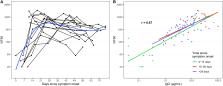
          We measured plasma and/or serum antibody responses to the receptor-binding domain (RBD) of the spike (S) protein of SARS-CoV-2 in 343 North American patients infected with SARS-CoV-2 (of which 93% required hospitalization) up to 122 days after symptom onset and compared them to responses in 1548 individuals whose blood samples were obtained prior to the pandemic. After setting seropositivity thresholds for perfect specificity (100%), we estimated sensitivities of 95% for IgG, 90% for IgA, and 81% for IgM for detecting infected individuals between 15 and 28 days after symptom onset. While the median time to seroconversion was nearly 12 days across all three isotypes tested, IgA and IgM antibodies against RBD were short-lived with median times to seroreversion of 71 and 49 days after symptom onset. In contrast, anti-RBD IgG responses decayed slowly through 90 days with only 3 seropositive individuals seroreverting within this time period. IgG antibodies to SARS-CoV-2 RBD were strongly correlated with anti-S neutralizing antibody titers, which demonstrated little to no decrease over 75 days since symptom onset. We observed no cross-reactivity of the SARS-CoV-2 RBD-targeted antibodies with other widely circulating coronaviruses (HKU1, 229 E, OC43, NL63). These data suggest that RBD-targeted antibodies are excellent markers of previous and recent infection, that differential isotype measurements can help distinguish between recent and older infections, and that IgG responses persist over the first few months after infection and are highly correlated with neutralizing antibodies.

          Related collections

          Most cited references19

          • Record: found
          • Abstract: found
          • Article: not found

          Clinical and immunological assessment of asymptomatic SARS-CoV-2 infections

          The clinical features and immune responses of asymptomatic individuals infected with severe acute respiratory syndrome coronavirus 2 (SARS-CoV-2) have not been well described. We studied 37 asymptomatic individuals in the Wanzhou District who were diagnosed with RT-PCR-confirmed SARS-CoV-2 infections but without any relevant clinical symptoms in the preceding 14 d and during hospitalization. Asymptomatic individuals were admitted to the government-designated Wanzhou People's Hospital for centralized isolation in accordance with policy1. The median duration of viral shedding in the asymptomatic group was 19 d (interquartile range (IQR), 15-26 d). The asymptomatic group had a significantly longer duration of viral shedding than the symptomatic group (log-rank P = 0.028). The virus-specific IgG levels in the asymptomatic group (median S/CO, 3.4; IQR, 1.6-10.7) were significantly lower (P = 0.005) relative to the symptomatic group (median S/CO, 20.5; IQR, 5.8-38.2) in the acute phase. Of asymptomatic individuals, 93.3% (28/30) and 81.1% (30/37) had reduction in IgG and neutralizing antibody levels, respectively, during the early convalescent phase, as compared to 96.8% (30/31) and 62.2% (23/37) of symptomatic patients. Forty percent of asymptomatic individuals became seronegative and 12.9% of the symptomatic group became negative for IgG in the early convalescent phase. In addition, asymptomatic individuals exhibited lower levels of 18 pro- and anti-inflammatory cytokines. These data suggest that asymptomatic individuals had a weaker immune response to SARS-CoV-2 infection. The reduction in IgG and neutralizing antibody levels in the early convalescent phase might have implications for immunity strategy and serological surveys.
            Bookmark
            • Record: found
            • Abstract: found
            • Article: not found

            Profiling Early Humoral Response to Diagnose Novel Coronavirus Disease (COVID-19)

            Abstract Background The emergence of coronavirus disease 2019 (COVID-19) is a major healthcare threat. The current method of detection involves a quantitative polymerase chain reaction (qPCR)–based technique, which identifies the viral nucleic acids when present in sufficient quantity. False-negative results can be achieved and failure to quarantine the infected patient would be a major setback in containing the viral transmission. We aim to describe the time kinetics of various antibodies produced against the 2019 novel coronavirus (SARS-CoV-2) and evaluate the potential of antibody testing to diagnose COVID-19. Methods The host humoral response against SARS-CoV-2, including IgA, IgM, and IgG response, was examined by using an ELISA-based assay on the recombinant viral nucleocapsid protein. 208 plasma samples were collected from 82 confirmed and 58 probable cases (qPCR negative but with typical manifestation). The diagnostic value of IgM was evaluated in this cohort. Results The median duration of IgM and IgA antibody detection was 5 (IQR, 3–6) days, while IgG was detected 14 (IQR, 10–18) days after symptom onset, with a positive rate of 85.4%, 92.7%, and 77.9%, respectively. In confirmed and probable cases, the positive rates of IgM antibodies were 75.6% and 93.1%, respectively. The detection efficiency by IgM ELISA is higher than that of qPCR after 5.5 days of symptom onset. The positive detection rate is significantly increased (98.6%) when combining IgM ELISA assay with PCR for each patient compared with a single qPCR test (51.9%). Conclusions The humoral response to SARS-CoV-2 can aid in the diagnosis of COVID-19, including subclinical cases.
              Bookmark
              • Record: found
              • Abstract: found
              • Article: not found

              Variation in False-Negative Rate of Reverse Transcriptase Polymerase Chain Reaction–Based SARS-CoV-2 Tests by Time Since Exposure

              Background: Tests for severe acute respiratory syndrome coronavirus 2 (SARS-CoV-2) based on reverse transcriptase polymerase chain reaction (RT-PCR) are being used to “rule out” infection among high-risk persons, such as exposed inpatients and health care workers. It is critical to understand how the predictive value of the test varies with time from exposure and symptom onset to avoid being falsely reassured by negative test results. Objective: To estimate the false-negative rate by day since infection. Design: Literature review and pooled analysis. Setting: 7 previously published studies providing data on RT-PCR performance by time since symptom onset or SARS-CoV-2 exposure using samples from the upper respiratory tract (n = 1330). Patients: A mix of inpatients and outpatients with SARS-CoV-2 infection. Measurements: A Bayesian hierarchical model was fitted to estimate the false-negative rate by day since exposure and symptom onset. Results: Over the 4 days of infection before the typical time of symptom onset (day 5), the probability of a false-negative result in an infected person decreases from 100% (95% CI, 100% to 100%) on day 1 to 67% (CI, 27% to 94%) on day 4. On the day of symptom onset, the median false-negative rate was 38% (CI, 18% to 65%). This decreased to 20% (CI, 12% to 30%) on day 8 (3 days after symptom onset) then began to increase again, from 21% (CI, 13% to 31%) on day 9 to 66% (CI, 54% to 77%) on day 21. Limitation: Imprecise estimates due to heterogeneity in the design of studies on which results were based. Conclusion: Care must be taken in interpreting RT-PCR tests for SARS-CoV-2 infection—particularly early in the course of infection—when using these results as a basis for removing precautions intended to prevent onward transmission. If clinical suspicion is high, infection should not be ruled out on the basis of RT-PCR alone, and the clinical and epidemiologic situation should be carefully considered. Primary Funding Source: National Institute of Allergy and Infectious Diseases, Johns Hopkins Health System, and U.S. Centers for Disease Control and Prevention.
                Bookmark

                Author and article information

                Contributors
                Role: Investigation
                Role: MethodologyRole: SupervisionRole: Validation
                Journal
                Sci Immunol
                Sci Immunol
                SciImmunol
                immunology
                Science Immunology
                American Association for the Advancement of Science
                2470-9468
                08 October 2020
                : 5
                : 52
                : eabe0367
                Affiliations
                [a ]Division of Infectious Diseases, Massachusetts General Hospital, Boston, MA, USA.
                [b ]Department of Medicine, Harvard Medical School, Boston, MA, USA.
                [c ]Department of Epidemiology, Johns Hopkins Bloomberg School of Public Health, Baltimore, MD, USA.
                [d ]Department of Pathology, Massachusetts General Hospital, Boston, MA, USA.
                [e ]Ragon Institute of MGH, MIT, and Harvard, Cambridge, MA, USA.
                [f ]Department of Microbiology, Harvard Medical School, Boston, MA, USA.
                [g ]Center for Virology and Vaccine Research, Beth Israel Deaconess Medical Center, Harvard Medical School, Boston, MA, USA.
                [h ]Department of Immunology and Infectious Diseases, Harvard T.H. Chan School of Public Health, Boston, MA, USA.
                [i ]Department of Pediatrics, Harvard Medical School, Boston, MA, USA.
                Author notes
                [*]

                contributed equally to this work.

                [#]

                contributed equally to this work.

                []Address correspondence to Richelle C. Charles, rcharles@ 123456mgh.harvard.edu
                Author information
                https://orcid.org/0000-0003-4334-1510
                https://orcid.org/0000-0003-1474-4871
                https://orcid.org/0000-0001-6920-8000
                https://orcid.org/0000-0002-1115-0894
                https://orcid.org/0000-0003-1888-8186
                https://orcid.org/0000-0002-5084-8523
                https://orcid.org/0000-0002-7954-673X
                https://orcid.org/0000-0002-2229-0622
                https://orcid.org/0000-0002-1269-1895
                https://orcid.org/0000-0002-3614-4976
                https://orcid.org/0000-0003-2307-3379
                https://orcid.org/0000-0002-7489-0232
                https://orcid.org/0000-0002-7888-4067
                https://orcid.org/0000-0002-2083-7153
                https://orcid.org/0000-0003-2948-630X
                https://orcid.org/0000-0002-0775-6623
                https://orcid.org/0000-0002-4387-9954
                https://orcid.org/0000-0002-0100-1684
                https://orcid.org/0000-0001-7138-4441
                https://orcid.org/0000-0002-3603-8110
                https://orcid.org/0000-0003-4558-0339
                https://orcid.org/0000-0002-7009-7253
                https://orcid.org/0000-0001-5127-4659
                https://orcid.org/0000-0003-3627-2553
                https://orcid.org/0000-0001-8662-9077
                https://orcid.org/0000-0002-7680-9215
                https://orcid.org/0000-0003-3642-5126
                Article
                abe0367
                10.1126/sciimmunol.abe0367
                7857394
                33033172
                abf1d408-7b59-4528-a4e7-74417f4b613c
                Copyright © 2020 The Authors, some rights reserved; exclusive licensee American Association for the Advancement of Science. No claim to original U.S. Government Works. Distributed under a Creative Commons Attribution License 4.0 (CC BY).

                This is an open-access article distributed under the terms of the Creative Commons Attribution license, which permits unrestricted use, distribution, and reproduction in any medium, provided the original work is properly cited.

                History
                : 28 July 2020
                : 05 October 2020
                Funding
                Funded by: doi http://dx.doi.org/10.13039/100000002, National Institutes of Health;
                Award ID: R01 AI146779
                Funded by: doi http://dx.doi.org/10.13039/100000002, National Institutes of Health;
                Award ID: T32 GM007753
                Funded by: doi http://dx.doi.org/10.13039/100000002, National Institutes of Health;
                Award ID: T32 GM007753
                Funded by: doi http://dx.doi.org/10.13039/100000002, National Institutes of Health;
                Award ID: T32 AI007245
                Funded by: doi http://dx.doi.org/10.13039/100000030, Centers for Disease Control and Prevention;
                Award ID: U01CK000490
                Funded by: doi http://dx.doi.org/10.13039/100000030, Centers for Disease Control and Prevention;
                Award ID: U01CK000490
                Funded by: doi http://dx.doi.org/10.13039/100000030, Centers for Disease Control and Prevention;
                Award ID: U01CK000490
                Funded by: doi http://dx.doi.org/10.13039/100000030, Centers for Disease Control and Prevention;
                Award ID: U01CK000490
                Funded by: doi http://dx.doi.org/10.13039/100000030, Centers for Disease Control and Prevention;
                Award ID: U01CK000490
                Funded by: doi http://dx.doi.org/10.13039/100000030, Centers for Disease Control and Prevention;
                Award ID: U01CK000490
                Funded by: doi http://dx.doi.org/10.13039/100000030, Centers for Disease Control and Prevention;
                Award ID: U01CK000490
                Funded by: MassCPR;
                Categories
                Reports
                Reports
                Reports
                Coronavirus

                Comments

                Comment on this article