50
views
0
recommends
+1 Recommend
0 collections
    0
    shares
      • Record: found
      • Abstract: found
      • Article: not found

      Isolation of quiescent and nonquiescent cells from yeast stationary-phase cultures

      research-article
      Bookmark
          There is no author summary for this article yet. Authors can add summaries to their articles on ScienceOpen to make them more accessible to a non-specialist audience.

          Abstract

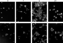
          Quiescence is the most common and, arguably, most poorly understood cell cycle state. This is in part because pure populations of quiescent cells are typically difficult to isolate. We report the isolation and characterization of quiescent and nonquiescent cells from stationary-phase (SP) yeast cultures by density-gradient centrifugation. Quiescent cells are dense, unbudded daughter cells formed after glucose exhaustion. They synchronously reenter the mitotic cell cycle, suggesting that they are in a G 0 state. Nonquiescent cells are less dense, heterogeneous, and composed of replicatively older, asynchronous cells that rapidly lose the ability to reproduce. Microscopic and flow cytometric analysis revealed that nonquiescent cells accumulate more reactive oxygen species than quiescent cells, and over 21 d, about half exhibit signs of apoptosis and necrosis. The ability to isolate both quiescent and nonquiescent yeast cells from SP cultures provides a novel, tractable experimental system for studies of quiescence, chronological and replicative aging, apoptosis, and the cell cycle.

          Related collections

          Most cited references46

          • Record: found
          • Abstract: found
          • Article: not found

          Genomic expression programs in the response of yeast cells to environmental changes.

          We explored genomic expression patterns in the yeast Saccharomyces cerevisiae responding to diverse environmental transitions. DNA microarrays were used to measure changes in transcript levels over time for almost every yeast gene, as cells responded to temperature shocks, hydrogen peroxide, the superoxide-generating drug menadione, the sulfhydryl-oxidizing agent diamide, the disulfide-reducing agent dithiothreitol, hyper- and hypo-osmotic shock, amino acid starvation, nitrogen source depletion, and progression into stationary phase. A large set of genes (approximately 900) showed a similar drastic response to almost all of these environmental changes. Additional features of the genomic responses were specialized for specific conditions. Promoter analysis and subsequent characterization of the responses of mutant strains implicated the transcription factors Yap1p, as well as Msn2p and Msn4p, in mediating specific features of the transcriptional response, while the identification of novel sequence elements provided clues to novel regulators. Physiological themes in the genomic responses to specific environmental stresses provided insights into the effects of those stresses on the cell.
            Bookmark
            • Record: found
            • Abstract: found
            • Article: not found

            Oxygen Stress: A Regulator of Apoptosis in Yeast

            Oxygen radicals are important components of metazoan apoptosis. We have found that apoptosis can be induced in the yeast Saccharomyces cerevisiae by depletion of glutathione or by low external doses of H2O2. Cycloheximide prevents apoptotic death revealing active participation of the cell. Yeast can also be triggered into apoptosis by a mutation in CDC48 or by expression of mammalian bax. In both cases, we show oxygen radicals to accumulate in the cell, whereas radical depletion or hypoxia prevents apoptosis. These results suggest that the generation of oxygen radicals is a key event in the ancestral apoptotic pathway and offer an explanation for the mechanism of bax-induced apoptosis in the absence of any established apoptotic gene in yeast.
              Bookmark
              • Record: found
              • Abstract: found
              • Article: not found

              A restriction point for control of normal animal cell proliferation.

              A B PARDEE (1974)
              This paper provides evidence that normal animal cells possess a unique regulatory mechanism to shift them between proliferative and quiescent states. Cells cease to increase in number under a diversity of suboptimal nutritional conditions, whereas a uniformity of metabolic changes follows these nutritional shifts. Evidence is given here that cells are put into the same quiescent state by each of these diverse blocks to proliferation and that cells escape at the same point in G(1) of the cell cycle when nutrition is restored. The name restriction point is proposed for the specific time in the cell cycle at which this critical release event occurs. The restriction point control is proposed to permit normal cells to retain viability by a shift to a minimal metabolism upon differentiation in vivo and in vitro when conditions are suboptimal for growth. Malignant cells are proposed to have lost their restriction point control. Hence, under very adverse conditions, as in the presence of antitumor agents, they stop randomly in their division cycle and die.
                Bookmark

                Author and article information

                Journal
                J Cell Biol
                JCB
                The Journal of Cell Biology
                The Rockefeller University Press
                0021-9525
                1540-8140
                3 July 2006
                : 174
                : 1
                : 89-100
                Affiliations
                [1 ]Department of Biology, [2 ]Department of Mathematics and Statistics, and [3 ]Department of Molecular Genetics and Microbiology, Health Sciences Center, University of New Mexico, Albuquerque, NM 87131
                [4 ]Institute of Molecular Biology, Biochemistry, and Microbiology, Karl-Franzens University, 8010 Graz, Austria
                [5 ]Department of Eukaryotic Microbiology, University of Groningen, Kerklaan 30, 9750 AA Haren, Netherlands
                Author notes

                Correspondence to Margaret Werner-Washburne: maggieww@ 123456unm.edu

                Article
                200604072
                10.1083/jcb.200604072
                2064167
                16818721
                860f71d1-bd59-4e05-ac86-74cbc8954940
                Copyright © 2006, The Rockefeller University Press
                History
                : 17 April 2006
                : 30 May 2006
                Categories
                Research Articles
                Article

                Cell biology
                Cell biology

                Comments

                Comment on this article