2
views
0
recommends
+1 Recommend
0 collections
    0
    shares
      • Record: found
      • Abstract: found
      • Article: found
      Is Open Access

      The Role of VP16 in the Life Cycle of Alphaherpesviruses

      review-article

      Read this article at

      Bookmark
          There is no author summary for this article yet. Authors can add summaries to their articles on ScienceOpen to make them more accessible to a non-specialist audience.

          Abstract

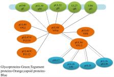
          The protein encoded by the UL48 gene of alphaherpesviruses is named VP16 or alpha-gene-transactivating factor (α-TIF). In the early stage of viral replication, VP16 is an important transactivator that can activate the transcription of viral immediate-early genes, and in the late stage of viral replication, VP16, as a tegument, is involved in viral assembly. This review will explain the mechanism of VP16 acting as α-TIF to activate the transcription of viral immediate-early genes, its role in the transition from viral latency to reactivation, and its effects on viral assembly and maturation. In addition, this review also provides new insights for further research on the life cycle of alphaherpesviruses and the role of VP16 in the viral life cycle.

          Related collections

          Most cited references150

          • Record: found
          • Abstract: found
          • Article: found
          Is Open Access

          Tegument Assembly and Secondary Envelopment of Alphaherpesviruses

          Alphaherpesviruses like herpes simplex virus are large DNA viruses characterized by their ability to establish lifelong latent infection in neurons. As for all herpesviruses, alphaherpesvirus virions contain a protein-rich layer called “tegument” that links the DNA-containing capsid to the glycoprotein-studded membrane envelope. Tegument proteins mediate a diverse range of functions during the virus lifecycle, including modulation of the host-cell environment immediately after entry, transport of virus capsids to the nucleus during infection, and wrapping of cytoplasmic capsids with membranes (secondary envelopment) during virion assembly. Eleven tegument proteins that are conserved across alphaherpesviruses have been implicated in the formation of the tegument layer or in secondary envelopment. Tegument is assembled via a dense network of interactions between tegument proteins, with the redundancy of these interactions making it challenging to determine the precise function of any specific tegument protein. However, recent studies have made great headway in defining the interactions between tegument proteins, conserved across alphaherpesviruses, which facilitate tegument assembly and secondary envelopment. We summarize these recent advances and review what remains to be learned about the molecular interactions required to assemble mature alphaherpesvirus virions following the release of capsids from infected cell nuclei.
            Bookmark
            • Record: found
            • Abstract: found
            • Article: not found

            The herpes simplex virus VP16-induced complex: the makings of a regulatory switch.

            When herpes simplex virus (HSV) infects human cells, it is able to enter two modes of infection: lytic and latent. A key activator of lytic infection is a virion protein called VP16, which, upon infection of a permissive cell, forms a transcriptional regulatory complex with two cellular proteins - the POU-domain transcription factor Oct-1 and the cell-proliferation factor HCF-1 - to activate transcription of the first set of expressed viral genes. This regulatory complex, called the VP16-induced complex, reveals mechanisms of combinatorial control of transcription. The activities of Oct-1 and HCF-1 - two important regulators of cellular gene expression and proliferation - illuminate strategies by which HSV might coexist with its host.
              Bookmark
              • Record: found
              • Abstract: found
              • Article: not found

              Transport and egress of herpes simplex virus in neurons.

              The mechanisms of axonal transport of the alphaherpesviruses, HSV and pseudorabies virus (PrV), in neuronal axons are of fundamental interest, particularly in comparison with other viruses, and offer potential sites for antiviral intervention or development of gene therapy vectors. These herpesviruses are transported rapidly along microtubules (MTs) in the retrograde direction from the axon terminus to the dorsal root ganglion and then anterogradely in the opposite direction. Retrograde transport follows fusion and deenvelopment of the viral capsid at the axonal membrane followed by loss of most of the tegument proteins and then binding of the capsid via one or more viral proteins (VPs) to the retrograde molecular motor dynein. The HSV capsid protein pUL35 has been shown to bind to the dynein light chain Tctex1 but is likely to be accompanied by additional dynein binding of an inner tegument protein. The mechanism of anterograde transport is much more controversial with different processes being claimed for PrV and HSV: separate transport of HSV capsid/tegument and glycoproteins versus PrV transport as an enveloped virion. The controversy has not been resolved despite application, in several laboratories, of confocal microscopy (CFM), real-time fluorescence with viruses dual labelled on capsid and glycoprotein, electron microscopy in situ and immuno-electron microscopy. Different processes for each virus seem counterintuitive although they are the most divergent in the alphaherpesvirus subfamily. Current hypotheses suggest that unenveloped HSV capsids complete assembly in the axonal growth cones and varicosities, whereas with PrV unenveloped capsids are only found travelling in a retrograde direction. John Wiley & Sons, Ltd
                Bookmark

                Author and article information

                Contributors
                Journal
                Front Microbiol
                Front Microbiol
                Front. Microbiol.
                Frontiers in Microbiology
                Frontiers Media S.A.
                1664-302X
                18 August 2020
                2020
                : 11
                : 1910
                Affiliations
                [1] 1Institute of Preventive Veterinary Medicine, Sichuan Agricultural University , Chengdu, China
                [2] 2Key Laboratory of Animal Disease and Human Health of Sichuan Province, Sichuan Agricultural University , Chengdu, China
                [3] 3Avian Disease Research Center, College of Veterinary Medicine, Sichuan Agricultural University , Chengdu, China
                Author notes

                Edited by: Akio Adachi, Kansai Medical University, Japan

                Reviewed by: Takayuki Murata, Fujita Health University, Japan; Evelyne Manet, UMR 5308 Centre International de Recherche en Infectiologie (CIRI), France

                *Correspondence: Mingshu Wang, mshwang@ 123456163.com

                These authors have contributed equally to this work and share first authorship

                This article was submitted to Virology, a section of the journal Frontiers in Microbiology

                Article
                10.3389/fmicb.2020.01910
                7461839
                33013729
                78aaba21-1ba1-475e-b798-3a3beda9100c
                Copyright © 2020 Fan, Wang, Cheng, Jia, Yang, Wu, Zhu, Zhao, Chen, Liu, Zhang, Ou, Mao, Gao, Sun, Wen, Liu, Yu, Zhang, Tian, Pan and Chen.

                This is an open-access article distributed under the terms of the Creative Commons Attribution License (CC BY). The use, distribution or reproduction in other forums is permitted, provided the original author(s) and the copyright owner(s) are credited and that the original publication in this journal is cited, in accordance with accepted academic practice. No use, distribution or reproduction is permitted which does not comply with these terms.

                History
                : 09 May 2020
                : 21 July 2020
                Page count
                Figures: 5, Tables: 2, Equations: 0, References: 156, Pages: 16, Words: 0
                Categories
                Microbiology
                Review

                Microbiology & Virology
                alphaherpesvirus,vp16,transcriptional activation,secondary envelopment,reactivation from latency

                Comments

                Comment on this article