- Record: found
- Abstract: found
- Article: not found
The determination of release from isolation of COVID-19 patients requires ultra-high sensitivity nucleic acid test technology
letter
Wanqiu Huang
1 ,
Dachuan Lin
2 ,
Cuini Wang
1 ,
Chaohui Bao
1 ,
Zhaoqi Zhang
4 ,
Xinchun Chen
2
,
3 ,
Zheng Zhang
3
,
** ,
Jian Huang
1
,
*
2 July 2020
Read this article at

There is no author summary for this article yet. Authors can add summaries to their articles on ScienceOpen to make them more accessible to a non-specialist audience.
Abstract
Dear Editor,
The prevention and control of SARS-CoV-2 has entered a critical period. Recent one
paper in this journal also discussed weather qualitative RT-PCR be used to determine
release from isolation of COVID-19 patients [1]. This issue is really important. Since
the outbreak of COVID-19 worldwide, discontinuation of isolation has been presenting
a dilemma of COVID-19, despite of the test-based strategy or the symptom-based strategy
[1]. The reason for the confusion is that nucleic acid testing presents false negative
based on qPCR technology, because of its low sensitivity 2, 3, 4. There are several
factors for false negative, including sample collection, preservation, transportation,
virus inactivation, nucleic acid extraction and technical sensitivity, among which
technical sensitivity and precise sampling are the most important quality control
measures to eliminate false negative.
It is well known that SARS-CoV-2 nucleic acid test is the main diagnostic method of
COVID-19. Recombinase polymerase amplification (RPA) is a new technology for testing
nucleic acid with some advantages of simple operation, fast speed and low cost based
on isothermal amplification. In our study, we developed an improved strategy, termed
as nestRPA (nest recombinase polymerase amplification), which could greatly improve
the sensitivity of nucleic acid detection for SARS-CoV-2 than RPA or qPCR.
Firstly, we designed eight sets of primers and probes for RPA on the conservation
regions of SARS-CoV-2 genes, in which some fragments were designed to span multiple
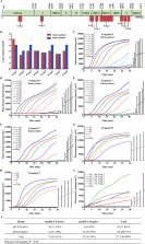
gene regions (Figure 1
A) which is one of the important technical tips. Through the two rounds of primer
screening, we found that the limit of detection (LOD) of 16 pairs of primers and 8
probes is quite different (Figure 1B), in which Fragment 1 against ORF1 gene had the
worst amplification efficiency. And Fragment 5 and 7 had the smallest LOD value, 300
and 500 copies/uL (Figure 1C to 1F), respectively.
Figure 1
Nucleic acid detection results using nestRPA. (A) The distribution of target fragments
on SARS-CoV-2 genome. (B) The LOD of optimum primer pairs from different gene regions.
(C) The sensitivity of outer primers for Fragment 5. (D) The sensitivity of inner
primers for Fragment 5. (E) The sensitivity of outer primers for Fragment 7. (F) The
sensitivity of inner primers for Fragment 7. (G) The sensitivity of nestRPA for Fragment
5. (H) The sensitivity of nestRPA for Fragment 7. (I) The five positive results of
four people returning to work by nestRPA. (J) Statistics of nucleic acid detection
results by nestRPA and qPCR assays for SARS-CoV-2. “*”, the statistical difference
of fluorescence intensity difference between test sample and blank control serves
as the criterion for judging the positive (p<0.05) of per reaction.
Figure 1
As far as we know, we firstly proposed the concept of nestRPA. The basic principles
of nestRPA are as follows: in nestRPA, the first amplification fragment of target
gene is amplified by outer primers, then a second fragment of target gene completely
within the first amplification fragment is amplified by inner primers. In order to
eliminate the influence of the fluorescence signal of enzymes, fluorescent probe is
not included in first RPA reaction which is another important technical tips. And
in the second RPA reaction, fluorescent probe will be added into reaction system.
Using nestRPA technology, we found that positive plasmid containing SARS-CoV-2 with
the concentration of 1 copy/ul could also be stably detected by Fragment 5 and Fragment
7 within 1-10 minutes (Figure 1G and 1H), suggesting that nestRPA technology indeed
performed very well for the detection of SARS-CoV-2 nucleic acid.
In order to promote the clinical application of nestRPA technology, we firstly collected
14 samples from 14 patients diagnosed as COVID-19, all of which SARS-CoV-2 nucleic
acid were positive using qPCR. The results of nestRPA assay showed that SARS-CoV-2
nucleic acid of these samples were 100% (14/14) positive. And then one positive sample
(Szt_P_002) with the lowest Cq-value was selected to test the sensitivity of nestRPA
technology. We found the detection result of Szt_P_002 sample was still positive after
11 times of 10-fold serial dilution by nestRPA assay, whilst after the fourth times
of the same dilution fold, the result by qPCR test has been negative. Secondly, 101
samples from 73 patients diagnosed as COVID-19 were collected, all of which had negative
results using qPCR, whilst 32.67% (33/101) of the samples were identified as by nestRPA
assay. Furthermore, we collected 25 samples from 8 re-positive patients who repeatedly
hospitalized suffering from COVID-19. Our results showed that 96.00% (24/25) of the
samples tested positive by nestRPA whilst only 24.00% (6/25) of the samples were confirmed
as positive by qPCR. These six samples with positive results by qPCR also had positive
results by nestRPA. Our detection results were basically consistent with the clinical
diagnosis results. Moreover, to explore whether there were asymptomatic patients with
SARS-CoV-2 nucleic acid positive in healthy population, we collected 32 nasal swabs
samples from those returning to work, all of which the SARS-CoV-2 nucleic acid detection
results were negative using qPCR. However, we found 12.50% (4/32) of the samples were
positive using Fragment 5 and/or Fragment 7 by nestRPA (Figure 1I), which was consistent
with those reported by other researchers [5]. Our results suggested that the ultra-sensitive
nucleic acid detection technique has important implications for early identification
of those asymptomatic carriers infected with SARS-CoV-2. Of course, in order to avoid
false positive results, the target sequence of positive amplification products was
100% detected by high-throughput sequencing. In summary, 36.18% (55/152) of the samples
with qPCR negative results were identified as positive by nestRPA technology in 172
clinical samples from 127 patients, which indicated the analytical sensitivity of
nestRPA assay is much better than that of qPCR (Figure 1J).
In addition, many experts of COVID-19 prevention and treatment clearly pointed out
that the inaccurate sample collection were also one of the important reasons for the
false negative result of SARS-CoV-2 nucleic acid 6, 7, 8. The most commonly sites
used as sampling are oropharynx and nasopharynx. The sample collectors should fix
the tongue with a spatula, and the sampling swab is used to scrape the cells from
tonsil recess and lateral wall when sampling from the oropharynx [9]. However, the
sample collectors were often fear of contagion with SARS-CoV-2. Under great infection
pressure, inaccurate sampling sites and inadequate sample volume will lead to false
negative test results. Therefore, it is helpful to reducing the false negative through
strict and normative operation of precise sampling with well protection for sample
collectors (Figure 2
).
Figure 2
Comparison of clinical sampling method and a protective sampling kit with light source.
(Left) Wrong sampling method; (Middle) Correct sampling method; (Right) protective
sampling kit with light source. This device is a protective oral-nasopharyngeal sampling
set with built-in light source, including 7 components: (1) LED inspection lamp handle;
(2) LED inspection light; (3) Disposable use of anti-droplet baffle; (4) U-shaped
slot; (5) Sterile swab; (6) Sampling hole; (7) Sterile tongue depressor.
Figure 2
Except for the technical sensitivity and precise sampling, we also need to pay more
attention for the quality control of sample preservation and transportation, virus
inactivation, nucleic acid extraction [10]. If all the links in the detection of SARS-CoV-2
nucleic acid could be strictly administrated, false negative could be completely eliminated,
and the discontinuation of isolation will no longer be a dilemma for us.
Author Contributions
All authors had full access to all the data in the study and take responsibility for
the integrity of the data and the accuracy of the data analysis. Jian Huang was responsible
for study concept and design. Zheng Zhang and Xinchun Chen were responsible for specimens
sampling. Wanqiu Huang and Dachuan Lin were responsible for the experiment and statistical
analysis. Wanqiu Huang, Dachuan Lin, Cuini Wang, Chaohui Bao and Zhaoqi Zhang were
responsible for the analysis of data. Wanqiu Huang and Jian Huang were responsible
for drafting the manuscript.
Declaration of Competing Interest
No authors declared any potential conflicts of interest.
Related collections
Most cited references10
- Record: found
- Abstract: found
- Article: not found
Antibody Detection and Dynamic Characteristics in Patients with COVID-19
Fei Xiang, Xiaorong Wang, Xinliang He … (2020)
- Record: found
- Abstract: found
- Article: found
False‐negative of RT‐PCR and prolonged nucleic acid conversion in COVID‐19: Rather than recurrence
Ai Tang Xiao, Yi Xin Tong, Sheng Zhang (2020)
- Record: found
- Abstract: found
- Article: not found