7
views
0
recommends
+1 Recommend
1 collections
    0
    shares
      • Record: found
      • Abstract: found
      • Article: found
      Is Open Access

      The fatty acid receptor CD36 promotes HCC progression through activating Src/PI3K/AKT axis-dependent aerobic glycolysis

      research-article

      Read this article at

      ScienceOpenPublisherPMC
      Bookmark
          There is no author summary for this article yet. Authors can add summaries to their articles on ScienceOpen to make them more accessible to a non-specialist audience.

          Abstract

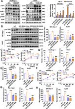
          Metabolic reprogramming is a new hallmark of cancer but it remains poorly defined in hepatocellular carcinogenesis (HCC). The fatty acid receptor CD36 is associated with both lipid and glucose metabolism in the liver. However, the role of CD36 in metabolic reprogramming in the progression of HCC still remains to be elucidated. In the present study, we found that CD36 is highly expressed in human HCC as compared with non-tumor hepatic tissue. CD36 overexpression promoted the proliferation, migration, invasion, and in vivo tumor growth of HCC cells, whereas silencing CD36 had the opposite effects. By analysis of cell metabolic phenotype, CD36 expression showed a positive association with extracellular acidification rate, a measure of glycolysis, instead of oxygen consumption rate. Further experiments verified that overexpression of CD36 resulted in increased glycolysis flux and lactic acid production. Mechanistically, CD36 induced mTOR-mediated oncogenic glycolysis via activation of Src/PI3K/AKT signaling axis. Pretreatment of HCC cells with PI3K/AKT/mTOR inhibitors largely blocked the tumor-promoting effect of CD36. Our findings suggest that CD36 exerts a stimulatory effect on HCC growth and metastasis, through mediating aerobic glycolysis by the Src/PI3K/AKT/mTOR signaling pathway.

          Related collections

          Most cited references42

          • Record: found
          • Abstract: found
          • Article: not found

          Global Cancer Statistics 2018: GLOBOCAN Estimates of Incidence and Mortality Worldwide for 36 Cancers in 185 Countries

          This article provides a status report on the global burden of cancer worldwide using the GLOBOCAN 2018 estimates of cancer incidence and mortality produced by the International Agency for Research on Cancer, with a focus on geographic variability across 20 world regions. There will be an estimated 18.1 million new cancer cases (17.0 million excluding nonmelanoma skin cancer) and 9.6 million cancer deaths (9.5 million excluding nonmelanoma skin cancer) in 2018. In both sexes combined, lung cancer is the most commonly diagnosed cancer (11.6% of the total cases) and the leading cause of cancer death (18.4% of the total cancer deaths), closely followed by female breast cancer (11.6%), prostate cancer (7.1%), and colorectal cancer (6.1%) for incidence and colorectal cancer (9.2%), stomach cancer (8.2%), and liver cancer (8.2%) for mortality. Lung cancer is the most frequent cancer and the leading cause of cancer death among males, followed by prostate and colorectal cancer (for incidence) and liver and stomach cancer (for mortality). Among females, breast cancer is the most commonly diagnosed cancer and the leading cause of cancer death, followed by colorectal and lung cancer (for incidence), and vice versa (for mortality); cervical cancer ranks fourth for both incidence and mortality. The most frequently diagnosed cancer and the leading cause of cancer death, however, substantially vary across countries and within each country depending on the degree of economic development and associated social and life style factors. It is noteworthy that high-quality cancer registry data, the basis for planning and implementing evidence-based cancer control programs, are not available in most low- and middle-income countries. The Global Initiative for Cancer Registry Development is an international partnership that supports better estimation, as well as the collection and use of local data, to prioritize and evaluate national cancer control efforts. CA: A Cancer Journal for Clinicians 2018;0:1-31. © 2018 American Cancer Society.
            Bookmark
            • Record: found
            • Abstract: found
            • Article: not found

            mTOR signaling in growth control and disease.

            The mechanistic target of rapamycin (mTOR) signaling pathway senses and integrates a variety of environmental cues to regulate organismal growth and homeostasis. The pathway regulates many major cellular processes and is implicated in an increasing number of pathological conditions, including cancer, obesity, type 2 diabetes, and neurodegeneration. Here, we review recent advances in our understanding of the mTOR pathway and its role in health, disease, and aging. We further discuss pharmacological approaches to treat human pathologies linked to mTOR deregulation. Copyright © 2012 Elsevier Inc. All rights reserved.
              Bookmark
              • Record: found
              • Abstract: found
              • Article: found

              Targeting metastasis-initiating cells through the fatty acid receptor CD36.

              The fact that the identity of the cells that initiate metastasis in most human cancers is unknown hampers the development of antimetastatic therapies. Here we describe a subpopulation of CD44(bright) cells in human oral carcinomas that do not overexpress mesenchymal genes, are slow-cycling, express high levels of the fatty acid receptor CD36 and lipid metabolism genes, and are unique in their ability to initiate metastasis. Palmitic acid or a high-fat diet specifically boosts the metastatic potential of CD36(+) metastasis-initiating cells in a CD36-dependent manner. The use of neutralizing antibodies to block CD36 causes almost complete inhibition of metastasis in immunodeficient or immunocompetent orthotopic mouse models of human oral cancer, with no side effects. Clinically, the presence of CD36(+) metastasis-initiating cells correlates with a poor prognosis for numerous types of carcinomas, and inhibition of CD36 also impairs metastasis, at least in human melanoma- and breast cancer-derived tumours. Together, our results indicate that metastasis-initiating cells particularly rely on dietary lipids to promote metastasis.
                Bookmark

                Author and article information

                Contributors
                yp_apple@yeah.net
                chenyaxi@cqmu.edu.cn
                Journal
                Cell Death Dis
                Cell Death Dis
                Cell Death & Disease
                Nature Publishing Group UK (London )
                2041-4889
                26 March 2021
                26 March 2021
                April 2021
                : 12
                : 4
                : 328
                Affiliations
                [1 ]GRID grid.203458.8, ISNI 0000 0000 8653 0555, Centre for Lipid Research and Key Laboratory of Molecular Biology for Infectious Diseases (Ministry of Education), Department of Infectious Diseases, Institute for Viral Hepatitis, The Second Affiliated Hospital, , Chongqing Medical University, ; 400016 Chongqing, China
                [2 ]GRID grid.83440.3b, ISNI 0000000121901201, John Moorhead Research Laboratory, Centre for Nephrology, University College London Medical School, , University College London, ; Royal Free Campus, London, NW3 2PF UK
                Author information
                http://orcid.org/0000-0001-5199-4971
                Article
                3596
                10.1038/s41419-021-03596-w
                7997878
                33771982
                69bf1fd6-c9dc-401b-a40e-1b7fb8fa8d7c
                © The Author(s) 2021

                Open Access This article is licensed under a Creative Commons Attribution 4.0 International License, which permits use, sharing, adaptation, distribution and reproduction in any medium or format, as long as you give appropriate credit to the original author(s) and the source, provide a link to the Creative Commons license, and indicate if changes were made. The images or other third party material in this article are included in the article’s Creative Commons license, unless indicated otherwise in a credit line to the material. If material is not included in the article’s Creative Commons license and your intended use is not permitted by statutory regulation or exceeds the permitted use, you will need to obtain permission directly from the copyright holder. To view a copy of this license, visit http://creativecommons.org/licenses/by/4.0/.

                History
                : 10 November 2020
                : 3 March 2021
                : 8 March 2021
                Funding
                Funded by: the Chongqing Research Program of Basic Research and Frontier Technology No.cstc2020jcyj-msxmX0205 the Science and Technology Project of Yuzhong District of Chongqing No.20190120
                Funded by: FundRef https://doi.org/10.13039/501100001809, National Natural Science Foundation of China (National Science Foundation of China);
                Award ID: 81873569
                Award Recipient :
                Funded by: the National Key R&D Program of China No.2018YFC1312700 the Chongqing Research Program of Basic Research and Frontier Technology No.cstc2020jcyj-zdxmX0007 the Science and Technology Research Program of Chongqing Municipal Education Commission No.KJZD-K201800401 the 111 Project No.D20028
                Categories
                Article
                Custom metadata
                © The Author(s) 2021

                Cell biology
                cancer metabolism,tumour biomarkers
                Cell biology
                cancer metabolism, tumour biomarkers

                Comments

                Comment on this article