11
views
0
recommends
+1 Recommend
0 collections
    0
    shares
      • Record: found
      • Abstract: found
      • Article: found
      Is Open Access

      Length to width ratio of the ductus venosus in simple screening for fetal congenital heart diseases in the second trimester

      research-article

      Read this article at

      Bookmark
          There is no author summary for this article yet. Authors can add summaries to their articles on ScienceOpen to make them more accessible to a non-specialist audience.

          Abstract

          Antenatal diagnosis of congenital heart disease (CHD) is still low even though screening was first introduced over 25 years ago. The purpose of our study was to determine the efficacy of a second-trimester prenatal ultrasonographic method of screening for CHD.

          From September 2012 to September 2013, the length and width of the fetal ductus venosus were measured sonographically in 1006 singleton fetuses, and the ratio of length to width was calculated. The accuracy of each fetal measurement and Doppler ultrasonography were determined. The standard fetal echocardiographic evaluations including 2-dimensional gray-scale imaging, color, and Doppler color flow mapping were performed. The transducer was aligned to the long axis of the fetal trunk to view the ductus venosus in its full length, including the inlet (isthmus) and outlet portions of the vessel. The diameters of the vessel inner wall and mid-point of the ductus venosus were measured using calipers. All scans and fetal measurements were conducted by a registered sonographer with more than 20 years of perinatal ultrasound screening experience.

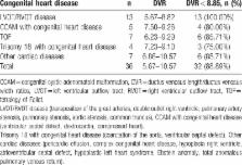
          Of the 1006 singleton fetuses between 19 +0 and 28 +6 weeks’ gestation, 36 had CHD. The ductus venosus length/width ratio (DVR) for the first CHD screening was extremely sensitive at 88.90%, with a specificity of 99.10% for the cardiac abnormalities included in this study. Chromosomal anomalies accompanied CHD in 0.4% (4/1006) of all cases and 11.11% (4/36) of the CHD cases.

          The DVR differed significantly between fetuses with CHD and normal fetuses during the second trimester. Careful assessment of the ratio should be a part of the sonographic examination of every fetus. In the case of a small DVR, advanced echocardiography and karyotype analysis should be performed. The ratio is a helpful tool for screening CHD abnormalities prenatally in the Chinese population.

          Related collections

          Most cited references28

          • Record: found
          • Abstract: not found
          • Article: not found

          American Society of Echocardiography guidelines and standards for performance of the fetal echocardiogram.

            Bookmark
            • Record: found
            • Abstract: found
            • Article: not found

            The examiner's ultrasound experience has a significant impact on the detection rate of congenital heart defects at the second-trimester fetal examination.

            To determine whether training and experience in performing ultrasound examinations are factors that influence the prenatal detection of congenital heart defects (CHDs) in a non-selected population, in order to evaluate and improve the current training program. All pregnant women who received a routine second-trimester ultrasound scan by a sonographer/midwife and delivered at our hospital between February 1991 and December 2001 were registered prospectively. Less experienced sonographer/midwives who had performed between 200 and 2000 routine examinations were compared with experienced sonographer/midwives who had carried out more than 2000 examinations. During the first 5 years of the study the heart structures obtained were registered in detail. Of 29,035 fetuses, 35/82 (43%) major CHDs were prenatally detected at the routine examination. The experienced sonographer/midwives obtained both the four-chamber view and the great arteries in 75%; the figure for the less experienced sonographer/midwives was 36% (P < 0.001). The differences in detecting major heart defects were 22/42 (52%) and 13/40 (32.5%), isolated CHDs 8/18 (44%) and 6/22 (27%) and CHDs with associated malformations 14/24 (58%) and 7/18 (39%), respectively. In both groups some CHDs with an abnormal four-chamber view were missed, although the experienced sonographer/midwives recognized significantly more of the abnormal views than did the less experienced sonographer/midwives (P = 0.002). Experience has a significant impact on the examination of the fetal heart and the prenatal detection rate of major CHDs. To avoid a relatively long learning curve, ultrasound education needs to intensify the teaching of the basic four-chamber view. The great arteries should be included after additional training. Those basic views of the fetal heart must be mastered before new views and advanced technology are added to the fetal heart examination. 2006 ISUOG. Published by John Wiley & Sons, Ltd.
              Bookmark
              • Record: found
              • Abstract: found
              • Article: not found

              Barriers to prenatal detection of congenital heart disease: a population-based study

              To evaluate the extent and determinants of missed prenatal detection of congenital heart disease (CHD) in a population-based setting.
                Bookmark

                Author and article information

                Journal
                Medicine (Baltimore)
                Medicine (Baltimore)
                MEDI
                Medicine
                Wolters Kluwer Health
                0025-7974
                1536-5964
                September 2016
                30 September 2016
                : 95
                : 39
                : e4928
                Affiliations
                [a ]Department of Biomedical Imaging and Radiological Sciences, School of Biomedical Science and Engineering, National Yang-Ming University
                [b ]Department of Obstetrics and Gynecology, Chung Shan Hospital, Taipei
                [c ]Department of Obstetrics and Gynecology, Hungchi Women & Children's Hospital, Taoyuan, Taiwan
                [d ]Department of Obstetrics and Gynecology, Changsha Anzhen Women Hospital, Changsha, China
                [e ]Fu-Jen Catholic University
                [f ]Department of Medical Research and Education, Cheng Hsin General Hospital, Taipei, Taiwan
                [g ]Department of Obstetrics and Gynecology, the 1st Affiliated Hospital, Jinan University, Guangzhou, China.
                Author notes
                []Correspondence: Ran-Chou Chen, Department of Biomedical Imaging and Radiological Sciences, School of Biomedical Science and Engineering, National Yang-Ming University, Taipei, Taiwan (e-mail: chenranchou@ 123456yahoo.com.tw ).
                Article
                04928
                10.1097/MD.0000000000004928
                5265924
                27684831
                63366afa-0446-4868-b5a8-769e9d365380
                Copyright © 2016 the Author(s). Published by Wolters Kluwer Health, Inc. All rights reserved.

                This is an open access article distributed under the Creative Commons Attribution-NoDerivatives License 4.0, which allows for redistribution, commercial and non-commercial, as long as it is passed along unchanged and in whole, with credit to the author. http://creativecommons.org/licenses/by-nd/4.0

                History
                : 8 April 2016
                : 5 August 2016
                : 18 August 2016
                Categories
                5600
                Research Article
                Diagnostic Accuracy Study
                Custom metadata
                TRUE

                congenital heart diseases,ductus venosus,prenatal
                congenital heart diseases, ductus venosus, prenatal

                Comments

                Comment on this article