7
views
0
recommends
+1 Recommend
1 collections
    0
    shares
      • Record: found
      • Abstract: found
      • Article: found
      Is Open Access

      Schizotypy-Related Magnetization of Cortex in Healthy Adolescence Is Colocated With Expression of Schizophrenia-Related Genes

      research-article

      Read this article at

      ScienceOpenPublisherPMC
      Bookmark
          There is no author summary for this article yet. Authors can add summaries to their articles on ScienceOpen to make them more accessible to a non-specialist audience.

          Abstract

          Background

          Genetic risk is thought to drive clinical variation on a spectrum of schizophrenia-like traits, but the underlying changes in brain structure that mechanistically link genomic variation to schizotypal experience and behavior are unclear.

          Methods

          We assessed schizotypy using a self-reported questionnaire and measured magnetization transfer as a putative microstructural magnetic resonance imaging marker of intracortical myelination in 68 brain regions in 248 healthy young people (14–25 years of age). We used normative adult brain gene expression data and partial least squares analysis to find the weighted gene expression pattern that was most colocated with the cortical map of schizotypy-related magnetization.

          Results

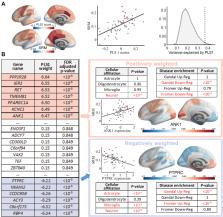
          Magnetization was significantly correlated with schizotypy in the bilateral posterior cingulate cortex and precuneus (and for disorganized schizotypy, also in medial prefrontal cortex; all false discovery rate–corrected p< .05), which are regions of the default mode network specialized for social and memory functions. The genes most positively weighted on the whole-genome expression map colocated with schizotypy-related magnetization were enriched for genes that were significantly downregulated in two prior case-control histological studies of brain gene expression in schizophrenia. Conversely, the most negatively weighted genes were enriched for genes that were transcriptionally upregulated in schizophrenia. Positively weighted (downregulated) genes were enriched for neuronal, specifically interneuronal, affiliations and coded a network of proteins comprising a few highly interactive “hubs” such as parvalbumin and calmodulin.

          Conclusions

          Microstructural magnetic resonance imaging maps of intracortical magnetization can be linked to both the behavioral traits of schizotypy and prior histological data on dysregulated gene expression in schizophrenia.

          Related collections

          Most cited references54

          • Record: found
          • Abstract: found
          • Article: not found

          BRAIN NETWORKS. Correlated gene expression supports synchronous activity in brain networks.

          During rest, brain activity is synchronized between different regions widely distributed throughout the brain, forming functional networks. However, the molecular mechanisms supporting functional connectivity remain undefined. We show that functional brain networks defined with resting-state functional magnetic resonance imaging can be recapitulated by using measures of correlated gene expression in a post mortem brain tissue data set. The set of 136 genes we identify is significantly enriched for ion channels. Polymorphisms in this set of genes significantly affect resting-state functional connectivity in a large sample of healthy adolescents. Expression levels of these genes are also significantly associated with axonal connectivity in the mouse. The results provide convergent, multimodal evidence that resting-state functional networks correlate with the orchestrated activity of dozens of genes linked to ion channel activity and synaptic function.
            Bookmark
            • Record: found
            • Abstract: found
            • Article: not found

            Hierarchy of transcriptomic specialization across human cortex captured by structural neuroimaging topography

            Hierarchy provides a unifying principle for the macroscale organization of anatomical and functional properties across primate cortex, yet microscale bases of specialization across human cortex are poorly understood. Anatomical hierarchy is conventionally informed by invasive tract-tracing measurements, creating a need for a principled proxy measure in humans. Moreover, cortex exhibits marked interareal variation in gene expression, yet organizing principles of cortical transcription remain unclear. We hypothesized that specialization of cortical microcircuitry involves hierarchical gradients of gene expression. We found that a noninvasive neuroimaging measure—MRI-derived T1w/T2w mapping—reliably indexes anatomical hierarchy, and captures the dominant pattern of transcriptional variation across human cortex. We found hierarchical gradients in expression profiles of genes related to microcircuit function, consistent with monkey microanatomy, and implicated in neuropsychiatric disorders. Our findings identify a hierarchical axis linking cortical transcription and anatomy, along which gradients of microscale properties may contribute to the macroscale specialization of cortical function.
              Bookmark
              • Record: found
              • Abstract: found
              • Article: not found

              Gene expression deficits in a subclass of GABA neurons in the prefrontal cortex of subjects with schizophrenia.

              Markers of inhibitory neurotransmission are altered in the prefrontal cortex (PFC) of subjects with schizophrenia, and several lines of evidence suggest that these alterations may be most prominent in the subset of GABA-containing neurons that express the calcium-binding protein, parvalbumin (PV). To test this hypothesis, we evaluated the expression of mRNAs for PV, another calcium-binding protein, calretinin (CR), and glutamic acid decarboxylase (GAD67) in postmortem brain specimens from 15 pairs of subjects with schizophrenia and matched control subjects using single- and dual-label in situ hybridization. Signal intensity for PV mRNA expression in PFC area 9 was significantly decreased in the subjects with schizophrenia, predominantly in layers III and IV. Analysis at the cellular level revealed that this decrease was attributable principally to a reduction in PV mRNA expression per neuron rather than by a decreased density of PV mRNA-positive neurons. In contrast, the same measures of CR mRNA expression were not altered in schizophrenia. These findings were confirmed by findings from cDNA microarray studies using different probes. Across the subjects with schizophrenia, the decrease in neuronal PV mRNA expression was highly associated (r = 0.84) with the decrease in the density of neurons containing detectable levels of GAD67 mRNA. Furthermore, simultaneous detection of PV and GAD67 mRNAs revealed that in subjects with schizophrenia only 55% of PV mRNA-positive neurons had detectable levels of GAD67 mRNA. Given the critical role that PV-containing GABA neurons appear to play in regulating the cognitive functions mediated by the PFC, the selective alterations in gene expression in these neurons may contribute to the cognitive deficits characteristic of schizophrenia.
                Bookmark

                Author and article information

                Contributors
                Journal
                Biol Psychiatry
                Biol. Psychiatry
                Biological Psychiatry
                Elsevier
                0006-3223
                1873-2402
                01 August 2020
                01 August 2020
                : 88
                : 3
                : 248-259
                Affiliations
                [a ]Department of Psychiatry, University of Cambridge, Cambridge, United Kingdom
                [b ]Alan Turing Institute, London, United Kingdom
                [c ]Research Department of Clinical, Educational and Health Psychology, London, United Kingdom
                [d ]Max Planck UCL Centre for Computational Psychiatry and Ageing Research, University College London, London, United Kingdom
                [e ]Wellcome Trust Centre for Neuroimaging, UCL Queen Square Institute of Neurology, University College London, London, United Kingdom
                [f ]School of Mathematical Sciences, Queen Mary University of London, London, United Kingdom
                [g ]Cambridgeshire and Peterborough NHS Foundation Trust, Huntingdon, United Kingdom
                Author notes
                []Address correspondence to Rafael Romero-Garcia Ph.D., University of Cambridge, Department of Psychiatry, Sir William Hardy Building, Downing Street, Cambridge CB2 3EB, United Kingdom. rr480@ 123456cam.ac.uk
                [1]

                PEV and ETB contributed equally to this work.

                Article
                S0006-3223(19)31922-5
                10.1016/j.biopsych.2019.12.005
                7369635
                32029217
                5c1611f5-4bba-4fdf-b607-776d49b12c8c
                Crown Copyright © 2019 Published by Elsevier Inc on behalf of Society of Biological Psychiatry.

                This is an open access article under the CC BY license (http://creativecommons.org/licenses/by/4.0/).

                History
                : 9 July 2019
                : 21 November 2019
                : 3 December 2019
                Categories
                Article

                Clinical Psychology & Psychiatry
                adolescence,allen human brain atlas,development,fast-spiking gabaergic interneurons,multiparameter mri mapping,myelination,schizophrenia

                Comments

                Comment on this article