4
views
0
recommends
+1 Recommend
0 collections
    0
    shares
      • Record: found
      • Abstract: found
      • Article: found
      Is Open Access

      A Tissue-Engineered Tracheobronchial In Vitro Co-Culture Model for Determining Epithelial Toxicological and Inflammatory Responses

      research-article

      Read this article at

      Bookmark
          There is no author summary for this article yet. Authors can add summaries to their articles on ScienceOpen to make them more accessible to a non-specialist audience.

          Abstract

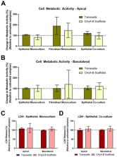
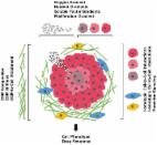
          Translation of novel inhalable therapies for respiratory diseases is hampered due to the lack of in vitro cell models that reflect the complexity of native tissue, resulting in many novel drugs and formulations failing to progress beyond preclinical assessments. The development of physiologically-representative tracheobronchial tissue analogues has the potential to improve the translation of new treatments by more accurately reflecting in vivo respiratory pharmacological and toxicological responses. Herein, advanced tissue-engineered collagen hyaluronic acid bilayered scaffolds (CHyA-B) previously developed within our group were used to evaluate bacterial and drug-induced toxicity and inflammation for the first time. Calu-3 bronchial epithelial cells and Wi38 lung fibroblasts were grown on either CHyA-B scaffolds (3D) or Transwell ® inserts (2D) under air liquid interface (ALI) conditions. Toxicological and inflammatory responses from epithelial monocultures and co-cultures grown in 2D or 3D were compared, using lipopolysaccharide (LPS) and bleomycin challenges to induce bacterial and drug responses in vitro. The 3D in vitro model exhibited significant epithelial barrier formation that was maintained upon introduction of co-culture conditions. Barrier integrity showed differential recovery in CHyA-B and Transwell ® epithelial cultures. Basolateral secretion of pro-inflammatory cytokines to bacterial challenge was found to be higher from cells grown in 3D compared to 2D. In addition, higher cytotoxicity and increased basolateral levels of cytokines were detected when epithelial cultures grown in 3D were challenged with bleomycin. CHyA-B scaffolds support the growth and differentiation of bronchial epithelial cells in a 3D co-culture model with different transepithelial resistance in comparison to the same co-cultures grown on Transwell ® inserts. Epithelial cultures in an extracellular matrix like environment show distinct responses in cytokine release and metabolic activity compared to 2D polarised models, which better mimic in vivo response to toxic and inflammatory stimuli offering an innovative in vitro platform for respiratory drug development.

          Related collections

          Most cited references79

          • Record: found
          • Abstract: found
          • Article: not found

          Modeling Physiological Events in 2D vs. 3D Cell Culture

          Cell culture has become an indispensable tool to help uncover fundamental biophysical and biomolecular mechanisms by which cells assemble into tissues and organs, how these tissues function, and how that function becomes disrupted in disease. Cell culture is now widely used in biomedical research, tissue engineering, regenerative medicine, and industrial practices. Although flat, two-dimensional (2D) cell culture has predominated, recent research has shifted toward culture using three-dimensional (3D) structures, and more realistic biochemical and biomechanical microenvironments. Nevertheless, in 3D cell culture, many challenges remain, including the tissue-tissue interface, the mechanical microenvironment, and the spatiotemporal distributions of oxygen, nutrients, and metabolic wastes. Here, we review 2D and 3D cell culture methods, discuss advantages and limitations of these techniques in modeling physiologically and pathologically relevant processes, and suggest directions for future research.
            Bookmark
            • Record: found
            • Abstract: found
            • Article: not found

            TEER measurement techniques for in vitro barrier model systems.

            Transepithelial/transendothelial electrical resistance (TEER) is a widely accepted quantitative technique to measure the integrity of tight junction dynamics in cell culture models of endothelial and epithelial monolayers. TEER values are strong indicators of the integrity of the cellular barriers before they are evaluated for transport of drugs or chemicals. TEER measurements can be performed in real time without cell damage and generally are based on measuring ohmic resistance or measuring impedance across a wide spectrum of frequencies. The measurements for various cell types have been reported with commercially available measurement systems and also with custom-built microfluidic implementations. Some of the barrier models that have been widely characterized using TEER include the blood-brain barrier (BBB), gastrointestinal (GI) tract, and pulmonary models. Variations in these values can arise due to factors such as temperature, medium formulation, and passage number of cells. The aim of this article is to review the different TEER measurement techniques and analyze their strengths and weaknesses, determine the significance of TEER in drug toxicity studies, examine the various in vitro models and microfluidic organs-on-chips implementations using TEER measurements in some widely studied barrier models (BBB, GI tract, and pulmonary), and discuss the various factors that can affect TEER measurements.
              Bookmark
              • Record: found
              • Abstract: found
              • Article: found
              Is Open Access

              Three-Dimensional in Vitro Cell Culture Models in Drug Discovery and Drug Repositioning

              Drug development is a lengthy and costly process that proceeds through several stages from target identification to lead discovery and optimization, preclinical validation and clinical trials culminating in approval for clinical use. An important step in this process is high-throughput screening (HTS) of small compound libraries for lead identification. Currently, the majority of cell-based HTS is being carried out on cultured cells propagated in two-dimensions (2D) on plastic surfaces optimized for tissue culture. At the same time, compelling evidence suggests that cells cultured in these non-physiological conditions are not representative of cells residing in the complex microenvironment of a tissue. This discrepancy is thought to be a significant contributor to the high failure rate in drug discovery, where only a low percentage of drugs investigated ever make it through the gamut of testing and approval to the market. Thus, three-dimensional (3D) cell culture technologies that more closely resemble in vivo cell environments are now being pursued with intensity as they are expected to accommodate better precision in drug discovery. Here we will review common approaches to 3D culture, discuss the significance of 3D cultures in drug resistance and drug repositioning and address some of the challenges of applying 3D cell cultures to high-throughput drug discovery.
                Bookmark

                Author and article information

                Contributors
                Role: Academic Editor
                Journal
                Biomedicines
                Biomedicines
                biomedicines
                Biomedicines
                MDPI
                2227-9059
                02 June 2021
                June 2021
                : 9
                : 6
                : 631
                Affiliations
                [1 ]School of Pharmacy and Biomolecular Sciences, RCSI University of Medicine and Health Sciences, D02 YN77 Dublin, Ireland; luissoriano@ 123456rcsi.com (L.S.); TehreemKhalid@ 123456rcsi.ie (T.K.); cianoleary@ 123456rcsi.ie (C.O.)
                [2 ]Tissue Engineering Research Group, Department of Anatomy and Regenerative Medicine, RCSI University of Medicine and Health Sciences, D02 YN77 Dublin, Ireland; fjobrien@ 123456rcsi.ie
                [3 ]SFI Centre for Research in Medical Devices (CÚRAM), RCSI University of Medicine and Health Sciences, D02 YN77 Dublin, Ireland
                [4 ]SFI Advanced Materials and Bioengineering Research (AMBER) Centre, RCSI University of Medicine and Health Sciences and Trinity College Dublin, D02 YN77 Dublin, Ireland
                [5 ]Trinity Centre for Biomedical Engineering, Trinity College Dublin, D02 PN40 Dublin, Ireland
                Author notes
                [* ]Correspondence: scryan@ 123456rcsi.ie
                Author information
                https://orcid.org/0000-0001-8624-9672
                https://orcid.org/0000-0001-6814-9731
                https://orcid.org/0000-0003-2030-8005
                https://orcid.org/0000-0002-9732-5703
                Article
                biomedicines-09-00631
                10.3390/biomedicines9060631
                8226664
                34199462
                532cc89d-b065-4ead-8d5c-e81c7eb6b69c
                © 2021 by the authors.

                Licensee MDPI, Basel, Switzerland. This article is an open access article distributed under the terms and conditions of the Creative Commons Attribution (CC BY) license ( https://creativecommons.org/licenses/by/4.0/).

                History
                : 04 May 2021
                : 29 May 2021
                Categories
                Article

                respiratory tissue engineering,collagen,toxicology,3d in vitro models,co-culture,epithelium,lipopolysaccharide,bleomycin,air-liquid interface

                Comments

                Comment on this article