25
views
0
recommends
+1 Recommend
0 collections
    0
    shares
      • Record: found
      • Abstract: found
      • Article: found
      Is Open Access

      Alterations of Regional Spontaneous Brain Activity and Gray Matter Volume in the Blind

      research-article

      Read this article at

      Bookmark
          There is no author summary for this article yet. Authors can add summaries to their articles on ScienceOpen to make them more accessible to a non-specialist audience.

          Abstract

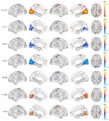
          Visual deprivation can induce alterations of regional spontaneous brain activity (RSBA). However, the effects of onset age of blindness on the RSBA and the association between the alterations of RSBA and brain structure are still unclear in the blind. In this study, we performed resting-state functional and structural magnetic resonance imaging on 50 sighted controls and 91 blind subjects (20 congenitally blind, 27 early blind, and 44 late blind individuals). Compared with the sighted control, we identified increased RSBA in the blind in primary and high-level visual areas and decreased RSBA in brain regions which are ascribed to sensorimotor and salience networks. In contrast, blind subjects exhibited significantly decreased gray matter volume (GMV) in the visual areas, while they exhibited significantly increased GMV in the sensorimotor areas. Moreover, the onset age of blindness was negatively correlated with the GMV of visual areas in blind subjects, whereas it exerted complex influences on the RSBA. Finally, significant negative correlations were shown between RSBA and GMV values. Our results demonstrated system-dependent, inverse alterations in RSBA and GMV after visual deprivation. Furthermore, the onset age of blindness has different effects on the reorganizations in RSBA and GMV.

          Related collections

          Most cited references43

          • Record: found
          • Abstract: found
          • Article: not found

          Connectivity-based parcellation of human cingulate cortex and its relation to functional specialization.

          Whole-brain neuroimaging studies have demonstrated regional variations in function within human cingulate cortex. At the same time, regional variations in cingulate anatomical connections have been found in animal models. It has, however, been difficult to estimate the relationship between connectivity and function throughout the whole cingulate cortex within the human brain. In this study, magnetic resonance diffusion tractography was used to investigate cingulate probabilistic connectivity in the human brain with two approaches. First, an algorithm was used to search for regional variations in the probabilistic connectivity profiles of all cingulate cortex voxels with the whole of the rest of the brain. Nine subregions with distinctive connectivity profiles were identified. It was possible to characterize several distinct areas in the dorsal cingulate sulcal region. Several distinct regions were also found in subgenual and perigenual cortex. Second, the probabilities of connection between cingulate cortex and 11 predefined target regions of interest were calculated. Cingulate voxels with a high probability of connection with the different targets formed separate clusters within cingulate cortex. Distinct connectivity fingerprints characterized the likelihood of connections between the extracingulate target regions and the nine cingulate subregions. Last, a meta-analysis of 171 functional studies reporting cingulate activation was performed. Seven different cognitive conditions were selected and peak activation coordinates were plotted to create maps of functional localization within the cingulate cortex. Regional functional specialization was found to be related to regional differences in probabilistic anatomical connectivity.
            Bookmark
            • Record: found
            • Abstract: found
            • Article: not found

            The origin of corticospinal projections from the premotor areas in the frontal lobe.

            We determined the origin of corticospinal neurons in the frontal lobe. These neurons were labeled by retrograde transport of tracers after injections into either the dorsolateral funiculus at the second cervical segment or the gray matter of the spinal cord throughout the cervical enlargement. Using retrograde transport of tracer from the arm area of the primary motor cortex, we defined the arm representation in each premotor area in another set of animals. We found that corticospinal projections to cervical segments of the spinal cord originate from the primary motor cortex and from the 6 premotor areas in the frontal lobe. These are the same premotor areas that project directly to the arm area of the primary motor cortex. The premotor areas are located in parts of cytoarchitectonic area 6 on the lateral surface and medial wall of the hemisphere, as well as in subfields of areas 23 and 24 in the cingulate sulcus. The total number of corticospinal neurons in the arm representations of the premotor areas equals or exceeds the total number in the arm representation of the primary motor cortex. The premotor areas collectively comprise more than 60% of the cortical area in the frontal lobe that projects to the spinal cord. Like the primary motor cortex, each of the premotor areas contains local regions that have a high density of corticospinal neurons. These observations indicate that a substantial component of the corticospinal system originates from the premotor areas in the frontal lobe. Each of the premotor areas has direct access to the spinal cord, and as a consequence, each has the potential to influence the generation and control of movement independently of the primary motor cortex. These findings raise serious questions about the utility of viewing the primary motor cortex as the "upper motoneuron" or "final common pathway" for the central control of movement.
              Bookmark
              • Record: found
              • Abstract: found
              • Article: not found

              Improved auditory spatial tuning in blind humans.

              Despite reports of improved auditory discrimination capabilities in blind humans and visually deprived animals, there is no general agreement as to the nature or pervasiveness of such compensatory sensory enhancements. Neuroimaging studies have pointed out differences in cerebral organization between blind and sighted humans, but the relationship between these altered cortical activation patterns and auditory sensory acuity remains unclear. Here we compare behavioural and electrophysiological indices of spatial tuning within central and peripheral auditory space in congenitally blind and normally sighted but blindfolded adults to test the hypothesis (raised by earlier studies of the effects of auditory deprivation on visual processing) that the effects of visual deprivation might be more pronounced for processing peripheral sounds. We find that blind participants displayed localization abilities that were superior to those of sighted controls, but only when attending to sounds in peripheral auditory space. Electrophysiological recordings obtained at the same time revealed sharper tuning of early spatial attention mechanisms in the blind subjects. Differences in the scalp distribution of brain electrical activity between the two groups suggest a compensatory reorganization of brain areas in the blind that may contribute to the improved spatial resolution for peripheral sound sources.
                Bookmark

                Author and article information

                Journal
                Neural Plast
                Neural Plast
                NP
                Neural Plasticity
                Hindawi Publishing Corporation
                2090-5904
                1687-5443
                2015
                19 October 2015
                : 2015
                : 141950
                Affiliations
                1Department of Radiology and Tianjin Key Laboratory of Functional Imaging, Tianjin Medical University General Hospital, Tianjin 300052, China
                2Brainnetome Center, Institute of Automation, Chinese Academy of Sciences, Beijing 100190, China
                Author notes

                Academic Editor: Lucas Pozzo-Miller

                Article
                10.1155/2015/141950
                4629052
                26568891
                399c924d-dff3-48c5-8e20-f885a18b6b4a
                Copyright © 2015 Aili Jiang et al.

                This is an open access article distributed under the Creative Commons Attribution License, which permits unrestricted use, distribution, and reproduction in any medium, provided the original work is properly cited.

                History
                : 23 December 2014
                : 20 March 2015
                : 25 March 2015
                Categories
                Research Article

                Neurosciences
                Neurosciences

                Comments

                Comment on this article