29
views
0
recommends
+1 Recommend
0 collections
    0
    shares
      • Record: found
      • Abstract: found
      • Article: found
      Is Open Access

      Altered White Matter Integrity in the Congenital and Late Blind People

      research-article

      Read this article at

      Bookmark
          There is no author summary for this article yet. Authors can add summaries to their articles on ScienceOpen to make them more accessible to a non-specialist audience.

          Abstract

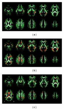
          The blind subjects have experienced a series of brain structural and functional alterations due to the visual deprivation. It remains unclear as to whether white matter changes differ between blind subjects with visual deprivation before and after a critical developmental period. The present study offered a direct comparison in changes of white matter fractional anisotropy (FA) between congenital blind (CB) and late blind (LB) individuals. Twenty CB, 21 LB (blindness onset after 18 years old), and 40 sight control (SC) subjects were recruited. Both the tract-based spatial statistics (TBSS) and voxel-based analysis (VBA) showed lower FA in the bilateral optic radiations in both blind groups, suggesting that the loss of white matter integrity was the prominent hallmark in the blind people. The LB group showed more extensive white matter impairment than the CB group, indicating the mechanisms of white matter FA changes are different between the CB and LB groups. Using a loose threshold, a trend of an increased FA was found in the bilateral corticospinal tracts in the LB but with a smaller spatial extent relative to the CB. These results suggest that white matter FA changes in the blind subjects are the reflection of multiple mechanisms, including the axonal degeneration, deafferentation, and plasticity.

          Related collections

          Most cited references50

          • Record: found
          • Abstract: found
          • Article: not found

          Water diffusion changes in Wallerian degeneration and their dependence on white matter architecture.

          This study investigates water diffusion changes in Wallerian degeneration. We measured indices derived from the diffusion tensor (DT) and T2-weighted signal intensities in the descending motor pathways of patients with small chronic lacunar infarcts of the posterior limb of the internal capsule on one side. We compared these measurements in the healthy and lesioned sides at different levels in the brainstem caudal to the primary lesion. We found that secondary white matter degeneration is revealed by a large reduction in diffusion anisotropy only in regions where fibers are arranged in isolated bundles of parallel fibers, such as in the cerebral peduncle. In regions where the degenerated pathway crosses other tracts, such as in the rostral pons, paradoxically there is almost no change in diffusion anisotropy, but a significant change in the measured orientation of fibers. The trace of the diffusion tensor is moderately increased in all affected regions. This allows one to differentiate secondary and primary fiber loss where the increase in trace is considerably higher. We show that DT-MRI is more sensitive than T2-weighted MRI in detecting Wallerian degeneration. Significant diffusion abnormalities are observed over the entire trajectory of the affected pathway in each patient. This finding suggests that mapping degenerated pathways noninvasively with DT-MRI is feasible. However, the interpretation of water diffusion data is complex and requires a priori information about anatomy and architecture of the pathway under investigation. In particular, our study shows that in regions where fibers cross, existing DT-MRI-based fiber tractography algorithms may lead to erroneous conclusion about brain connectivity.
            Bookmark
            • Record: found
            • Abstract: found
            • Article: not found

            Early 'visual' cortex activation correlates with superior verbal memory performance in the blind.

            The visual cortex may be more modifiable than previously considered. Using functional magnetic resonance imaging (fMRI) in ten congenitally blind human participants, we found robust occipital activation during a verbal-memory task (in the absence of any sensory input), as well as during verb generation and Braille reading. We also found evidence for reorganization and specialization of the occipital cortex, along the anterior-posterior axis. Whereas anterior regions showed preference for Braille, posterior regions (including V1) showed preference for verbal-memory and verb generation (which both require memory of verbal material). No such occipital activation was found in sighted subjects. This difference between the groups was mirrored by superior performance of the blind in various verbal-memory tasks. Moreover, the magnitude of V1 activation during the verbal-memory condition was highly correlated with the blind individual's abilities in a variety of verbal-memory tests, suggesting that the additional occipital activation may have a functional role.
              Bookmark
              • Record: found
              • Abstract: found
              • Article: not found

              Fractional anisotropy of water diffusion in cerebral white matter across the lifespan.

              Determining the time of peak of cerebral maturation is vital for our understanding of when cerebral maturation ceases and the cerebral degeneration in healthy aging begins. We carefully mapped changes in fractional anisotropy (FA) of water diffusion for eleven major cerebral white matter tracts in a large group (831) of healthy human subjects aged 11-90. FA is a neuroimaging index of micro-structural white matter integrity, sensitive to age-related changes in cerebral myelin levels, measured using diffusion tensor imaging. The average FA values of cerebral white matter (WM) reached peak at the age 32 ± 6 years. FA measurements for all but one major cortical white matter tract (cortico-spinal) reached peaks between 23 and 39 years of age. The maturation rates, prior to age-of-peak were significantly correlated (r=0.74; p=0.01) with the rates of decline, past age-of-peak. Regional analysis of corpus callosum (CC) showed that thinly-myelinated, densely packed fibers in the genu, that connect pre-frontal areas, maturated later and showed higher decline in aging than the more thickly myelinated motor and sensory areas in the body and splenium of CC. Our findings can be summarized as: associative, cerebral WM tracts that reach their peak FA values later in life also show progressively higher age-related decline than earlier maturing motor and sensory tracts. These findings carry multiple and diverse implications for both theoretical studies of the neurobiology of maturation and aging and for the clinical studies of neuropsychiatric disorders. Copyright © 2010 Elsevier Inc. All rights reserved.
                Bookmark

                Author and article information

                Journal
                Neural Plast
                Neural Plast
                NP
                Neural Plasticity
                Hindawi Publishing Corporation
                2090-5904
                1687-5443
                2013
                18 April 2013
                : 2013
                : 128236
                Affiliations
                1Department of Radiology, Tianjin Medical University General Hospital, Tianjin 300052, China
                2Brainnetome Center, Institute of Automation, Chinese Academy of Sciences, Beijing 100190, China
                3National Laboratory of Pattern Recognition, Institute of Automation, Chinese Academy of Sciences, Beijing 100190, China
                Author notes

                Academic Editor: Hao Lei

                Article
                10.1155/2013/128236
                3654351
                23710371
                fb7533e7-5252-44f2-bc72-665004545ae0
                Copyright © 2013 Dawei Wang et al.

                This is an open access article distributed under the Creative Commons Attribution License, which permits unrestricted use, distribution, and reproduction in any medium, provided the original work is properly cited.

                History
                : 5 March 2013
                : 1 April 2013
                Categories
                Research Article

                Neurosciences
                Neurosciences

                Comments

                Comment on this article