8
views
0
recommends
+1 Recommend
0 collections
    0
    shares
      • Record: found
      • Abstract: found
      • Article: found
      Is Open Access

      Improvement of Symptoms after Lymphaticovenous Anastomosis in Patients with Abdominal Wall Lymphedema

      research-article

      Read this article at

      Bookmark
          There is no author summary for this article yet. Authors can add summaries to their articles on ScienceOpen to make them more accessible to a non-specialist audience.

          Summary:

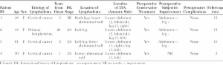
          At our institution, we performed a lymphaticovenous anastomosis in patients with primary or secondary abdominal lymphedema. Patients report good outcomes and feel relieved of their complaints. To obtain good results, it is important to have decent knowledge on the anatomical state of the lymphatic system. In general, the lymphatic system of the lower abdomen can be compared with the system of the upper legs. According to our current case results, the abdominal area might be susceptible to lymphaticovenous anastomosis procedure. Further research should be performed to confirm the effect of the intervention and the imaging techniques to monitor the improvements.

          Related collections

          Most cited references4

          • Record: found
          • Abstract: found
          • Article: not found

          Assessment and follow-up of patency after lymphovenous microsurgery for treatment of secondary lymphedema in external male genital organs.

          Secondary lymphedema of external male genital organs is a frequent complication of pelvic radical surgery following pelvic lymphadenectomy. Microsurgical lymphovenous anastomoses are usually performed using only the superficial scrotal lymphatics, excluding testicular lymphatic drainage. We have experimented using a new microsurgical technique based on lymphovenous anastomosis between the collectors of the spermatic funiculus and the veins of the pampiniform plexus, allowing testicular lymphatic drainage. The study included 11 patients with external genital organ lymphedema, five of whom were subjected to microsurgical lymphovenous derivation. At 3, 6, and 12 mo after surgery, the patency of lymphovenous anastomoses was assessed by noninvasive lymphography using indocyanine green fluorescence images obtained with the Photodynamic Eye (PDE) infrared camera system (Hamamatsu Photonics K.K., Hamamatsu, Japan). Progressive improvement of clinical conditions was assessed both by patients' self evaluation and by objective clinical follow-up based on: (1) PDE lymphography, (2) tomography of the pubic area, (3) recovery of the soft consistency of the scrotal tissue, (4) recovery of the scrotal skin normochromic aspect, (5) absence of pain, and (6) disappearance of edema with evident reduction of the scrotal and penile dimensions and normal palpability of the testis. The present study shows that lymphovenous anastomosis is a valuable method of resolving the edematous condition. The indocyanine green approach for lymphangiography is a very supportive method during follow-up because, with the least invasive approach, it is possible to ascertain the complete patency of the anastomosis, to confirm its localization, and to assess its lymphatic drainage.
            Bookmark
            • Record: found
            • Abstract: found
            • Article: not found

            Anatomy of the superficial lymphatics of the abdominal wall and the upper thigh and its implications in lymphatic microsurgery.

            The recent advent in the surgical treatment of lymphedema necessitates a more detailed understanding of the anatomy of the lymphatic system. Lymphovenous anastomosis (LVA) requires a precise knowledge of the anatomy of the superficial lymphatic collectors in relation to the superficial veins. In vascularized lymph node transfer (VLNT), donor site lymphatic function must be preserved.
              Bookmark
              • Record: found
              • Abstract: found
              • Article: not found

              Using indocyanine green fluorescent lymphography and lymphatic-venous anastomosis for cancer-related lymphedema.

              Advances in cancer therapy have increased the importance of improvement of quality of life after cancer survival. Cancer-related lymphedema or secondary lymphedema that occurs after lymph node dissection in resection of tumors of abdominal visceral organs can impair quality of life. However, standard curative treatment for secondary lymphedema has not been established. This may be due to the lack of a method for early diagnosis of lymphedema, and because of selection of conservative treatment such as compression therapy to delay edema progression in many cases. To develop a curative approach, we have performed definite diagnosis of early-stage lymphedema using magnetic resonance imaging and an indocyanine green fluorescent lymphography, followed by surgical treatment with lymphatic-venous anastomosis using supermicrosurgery. Herein, we report the first case of secondary lymphedema in which we performed early diagnosis and surgery using these techniques and achieved an almost complete cure of lymphedema. We suggest that early diagnostic imaging and early microsurgery is the key of lymphedema treatment.
                Bookmark

                Author and article information

                Journal
                Plast Reconstr Surg Glob Open
                Plast Reconstr Surg Glob Open
                GOX
                Plastic and Reconstructive Surgery Global Open
                Wolters Kluwer Health
                2169-7574
                12 March 2018
                March 2018
                : 6
                : 3
                : e1710
                Affiliations
                From the [* ]Department of General Surgery, Maastricht University Medical Center, Maastricht, The Netherlands; and []Department of Plastic and Reconstructive Surgery, Maastricht University Medical Center, Maastricht, The Netherlands.
                Author notes
                Shan Shan Qiu, MD, PhD, Department of Plastic and Reconstructive Surgery, Maastricht University Medical Center, P. Debyelaan 25. 6229 HX, Maastricht, The Netherlands, E-mail: shanshan.qiushao@ 123456mumc.nl
                Article
                00022
                10.1097/GOX.0000000000001710
                5908492
                2bafc451-b81c-4f70-ab77-e68b05cd76c8
                Copyright © 2018 The Authors. Published by Wolters Kluwer Health, Inc. on behalf of The American Society of Plastic Surgeons.

                This is an open-access article distributed under the terms of the Creative Commons Attribution-Non Commercial-No Derivatives License 4.0 (CCBY-NC-ND), where it is permissible to download and share the work provided it is properly cited. The work cannot be changed in any way or used commercially without permission from the journal.

                History
                : 21 July 2017
                : 19 December 2017
                Categories
                Ideas and Innovations
                Custom metadata
                TRUE

                Comments

                Comment on this article