82
views
0
recommends
+1 Recommend
0 collections
    0
    shares
      • Record: found
      • Abstract: found
      • Article: found
      Is Open Access

      The auditory and non-auditory brain areas involved in tinnitus. An emergent property of multiple parallel overlapping subnetworks

      review-article

      Read this article at

      Bookmark
          There is no author summary for this article yet. Authors can add summaries to their articles on ScienceOpen to make them more accessible to a non-specialist audience.

          Abstract

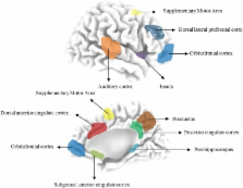
          Tinnitus is the perception of a sound in the absence of an external sound source. It is characterized by sensory components such as the perceived loudness, the lateralization, the tinnitus type (pure tone, noise-like) and associated emotional components, such as distress and mood changes. Source localization of quantitative electroencephalography (qEEG) data demonstrate the involvement of auditory brain areas as well as several non-auditory brain areas such as the anterior cingulate cortex (dorsal and subgenual), auditory cortex (primary and secondary), dorsal lateral prefrontal cortex, insula, supplementary motor area, orbitofrontal cortex (including the inferior frontal gyrus), parahippocampus, posterior cingulate cortex and the precuneus, in different aspects of tinnitus. Explaining these non-auditory brain areas as constituents of separable subnetworks, each reflecting a specific aspect of the tinnitus percept increases the explanatory power of the non-auditory brain areas involvement in tinnitus. Thus, the unified percept of tinnitus can be considered an emergent property of multiple parallel dynamically changing and partially overlapping subnetworks, each with a specific spontaneous oscillatory pattern and functional connectivity signature.

          Related collections

          Most cited references86

          • Record: found
          • Abstract: found
          • Article: not found

          Interoception: the sense of the physiological condition of the body.

          Converging evidence indicates that primates have a distinct cortical image of homeostatic afferent activity that reflects all aspects of the physiological condition of all tissues of the body. This interoceptive system, associated with autonomic motor control, is distinct from the exteroceptive system (cutaneous mechanoreception and proprioception) that guides somatic motor activity. The primary interoceptive representation in the dorsal posterior insula engenders distinct highly resolved feelings from the body that include pain, temperature, itch, sensual touch, muscular and visceral sensations, vasomotor activity, hunger, thirst, and 'air hunger'. In humans, a meta-representation of the primary interoceptive activity is engendered in the right anterior insula, which seems to provide the basis for the subjective image of the material self as a feeling (sentient) entity, that is, emotional awareness.
            Bookmark
            • Record: found
            • Abstract: found
            • Article: not found

            Neurobiology of emotion perception I: The neural basis of normal emotion perception.

            There is at present limited understanding of the neurobiological basis of the different processes underlying emotion perception. We have aimed to identify potential neural correlates of three processes suggested by appraisalist theories as important for emotion perception: 1) the identification of the emotional significance of a stimulus; 2) the production of an affective state in response to 1; and 3) the regulation of the affective state. In a critical review, we have examined findings from recent animal, human lesion, and functional neuroimaging studies. Findings from these studies indicate that these processes may be dependent upon the functioning of two neural systems: a ventral system, including the amygdala, insula, ventral striatum, and ventral regions of the anterior cingulate gyrus and prefrontal cortex, predominantly important for processes 1 and 2 and automatic regulation of emotional responses; and a dorsal system, including the hippocampus and dorsal regions of anterior cingulate gyrus and prefrontal cortex, predominantly important for process 3. We suggest that the extent to which a stimulus is identified as emotive and is associated with the production of an affective state may be dependent upon levels of activity within these two neural systems.
              Bookmark
              • Record: found
              • Abstract: found
              • Article: not found

              Conscious, preconscious, and subliminal processing: a testable taxonomy.

              Of the many brain events evoked by a visual stimulus, which are specifically associated with conscious perception, and which merely reflect non-conscious processing? Several recent neuroimaging studies have contrasted conscious and non-conscious visual processing, but their results appear inconsistent. Some support a correlation of conscious perception with early occipital events, others with late parieto-frontal activity. Here we attempt to make sense of these dissenting results. On the basis of the global neuronal workspace hypothesis, we propose a taxonomy that distinguishes between vigilance and access to conscious report, as well as between subliminal, preconscious and conscious processing. We suggest that these distinctions map onto different neural mechanisms, and that conscious perception is systematically associated with surges of parieto-frontal activity causing top-down amplification.
                Bookmark

                Author and article information

                Journal
                Front Syst Neurosci
                Front Syst Neurosci
                Front. Syst. Neurosci.
                Frontiers in Systems Neuroscience
                Frontiers Media S.A.
                1662-5137
                08 May 2012
                2012
                : 6
                : 31
                Affiliations
                simpleBrai2n, TRI and Department of Neurosurgery, University Hospital Antwerp Antwerp, Belgium
                Author notes

                Edited by: Jos J. Eggermont, University of Calgary, Canada

                Reviewed by: Dave Langers, University Medical Center Groningen, Netherlands; Winfried Schlee, Ulm University, Germany

                *Correspondence: Sven Vanneste, Brai 2n, TRI and Department of Neurosurgery, University Hospital Antwerp, Wilrijkstraat 10, 2650 Edegem, Belgium. e-mail: sven.vanneste@ 123456ua.ac.be
                Article
                10.3389/fnsys.2012.00031
                3347475
                22586375
                fad72d63-90a0-4798-893c-a7ed4235982a
                Copyright © 2012 Vanneste and De Ridder.

                This is an open-access article distributed under the terms of the Creative Commons Attribution Non Commercial License, which permits non-commercial use, distribution, and reproduction in other forums, provided the original authors and source are credited.

                History
                : 28 December 2011
                : 13 April 2012
                Page count
                Figures: 1, Tables: 1, Equations: 0, References: 110, Pages: 9, Words: 7807
                Categories
                Neuroscience
                Review Article

                Neurosciences
                emergent property,non-auditory brain areas,multiple parallel overlapping subnetworks,eeg

                Comments

                Comment on this article