0
views
0
recommends
+1 Recommend
0 collections
    0
    shares
      • Record: found
      • Abstract: found
      • Article: found
      Is Open Access

      Evaluation of the quality and the productivity of neuroradiological reading of multiple sclerosis follow-up MRI scans using an intelligent automation software

      research-article

      Read this article at

      Bookmark
          There is no author summary for this article yet. Authors can add summaries to their articles on ScienceOpen to make them more accessible to a non-specialist audience.

          Abstract

          Purpose

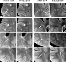
          The assessment of multiple sclerosis (MS) lesions on follow-up magnetic resonance imaging (MRI) is tedious, time-consuming, and error-prone. Automation of low-level tasks could enhance the radiologist in this work. We evaluate the intelligent automation software Jazz in a blinded three centers study, for the assessment of new, slowly expanding, and contrast-enhancing MS lesions.

          Methods

          In three separate centers, 117 MS follow-up MRIs were blindly analyzed on fluid attenuated inversion recovery (FLAIR), pre- and post-gadolinium T1-weighted images using Jazz by 2 neuroradiologists in each center. The reading time was recorded. The ground truth was defined in a second reading by side-by-side comparison of both reports from Jazz and the standard clinical report. The number of described new, slowly expanding, and contrast-enhancing lesions described with Jazz was compared to the lesions described in the standard clinical report.

          Results

          A total of 96 new lesions from 41 patients and 162 slowly expanding lesions (SELs) from 61 patients were described in the ground truth reading. A significantly larger number of new lesions were described using Jazz compared to the standard clinical report (63 versus 24). No SELs were reported in the standard clinical report, while 95 SELs were reported on average using Jazz. A total of 4 new contrast-enhancing lesions were found in all reports. The reading with Jazz was very time efficient, taking on average 2min33s ± 1min0s per case. Overall inter-reader agreement for new lesions between the readers using Jazz was moderate for new lesions (Cohen kappa = 0.5) and slight for SELs (0.08).

          Conclusion

          The quality and the productivity of neuroradiological reading of MS follow-up MRI scans can be significantly improved using the dedicated software Jazz.

          Related collections

          Most cited references44

          • Record: found
          • Abstract: found
          • Article: not found

          Visual attention: the past 25 years.

          This review focuses on covert attention and how it alters early vision. I explain why attention is considered a selective process, the constructs of covert attention, spatial endogenous and exogenous attention, and feature-based attention. I explain how in the last 25 years research on attention has characterized the effects of covert attention on spatial filters and how attention influences the selection of stimuli of interest. This review includes the effects of spatial attention on discriminability and appearance in tasks mediated by contrast sensitivity and spatial resolution; the effects of feature-based attention on basic visual processes, and a comparison of the effects of spatial and feature-based attention. The emphasis of this review is on psychophysical studies, but relevant electrophysiological and neuroimaging studies and models regarding how and where neuronal responses are modulated are also discussed. Copyright © 2011 Elsevier Ltd. All rights reserved.
            Bookmark
            • Record: found
            • Abstract: found
            • Article: not found

            Multiple sclerosis

            Multiple sclerosis continues to be a challenging and disabling condition but there is now greater understanding of the underlying genetic and environmental factors that drive the condition, including low vitamin D levels, cigarette smoking, and obesity. Early and accurate diagnosis is crucial and is supported by diagnostic criteria, incorporating imaging and spinal fluid abnormalities for those presenting with a clinically isolated syndrome. Importantly, there is an extensive therapeutic armamentarium, both oral and by infusion, for those with the relapsing remitting form of the disease. Careful consideration is required when choosing the correct treatment, balancing the side-effect profile with efficacy and escalating as clinically appropriate. This move towards more personalised medicine is supported by a clinical guideline published in 2018. Finally, a comprehensive management programme is strongly recommended for all patients with multiple sclerosis, enhancing health-related quality of life through advocating wellness, addressing aggravating factors, and managing comorbidities. The greatest remaining challenge for multiple sclerosis is the development of treatments incorporating neuroprotection and remyelination to treat and ultimately prevent the disabling, progressive forms of the condition.
              Bookmark
              • Record: found
              • Abstract: found
              • Article: not found

              Siponimod versus placebo in secondary progressive multiple sclerosis (EXPAND): a double-blind, randomised, phase 3 study

              No treatment has consistently shown efficacy in slowing disability progression in patients with secondary progressive multiple sclerosis (SPMS). We assessed the effect of siponimod, a selective sphingosine 1-phosphate (S1P) receptor1,5 modulator, on disability progression in patients with SPMS.
                Bookmark

                Author and article information

                Contributors
                christian@ai-medical.ch
                Journal
                Neuroradiology
                Neuroradiology
                Neuroradiology
                Springer Berlin Heidelberg (Berlin/Heidelberg )
                0028-3940
                1432-1920
                24 January 2024
                24 January 2024
                2024
                : 66
                : 3
                : 361-369
                Affiliations
                [1 ]AI Medical AG, Goldhaldenstr 22a, 8702 Zollikon, Switzerland
                [2 ]University of Zürich, ( https://ror.org/02crff812) Zürich, Switzerland
                [3 ]Department of Neuroradiology, Clinical Neuroscience Center, University Hospital Zürich, University of Zürich, ( https://ror.org/02crff812) Zürich, Switzerland
                [4 ]GRID grid.50550.35, ISNI 0000 0001 2175 4109, Department of Radiology, APHP, , Hôpitaux Raymond-Poincaré & Ambroise Paré, ; Paris, France
                [5 ]GRID grid.460789.4, ISNI 0000 0004 4910 6535, Laboratoire d’imagerie Biomédicale Multimodale (BioMaps), , Université Paris-Saclay, CEA, CNRS, Inserm, Service Hopsitalier Frédéric Joliot, ; Orsay, France
                [6 ]Stanford University, ( https://ror.org/00f54p054) Stanford, USA
                [7 ]Faculty of Medicine, University of Novi Sad, ( https://ror.org/00xa57a59) Novi Sad, Serbia
                [8 ]University Hospital Basel and University of Basel, ( https://ror.org/02s6k3f65) Basel, Switzerland
                [9 ]Department of Neurology, Poissy-Saint-Germain-en-Laye Hospital, Poissy, France
                [10 ]CRC SEP IDF Ouest, Poissy-Garches, France
                [11 ]GRID grid.168010.e, ISNI 0000000419368956, Department of Neurology & Neurological Sciences, , Stanford University School of Medicine, ; Stanford, CA USA
                [12 ]GRID grid.240145.6, ISNI 0000 0001 2291 4776, MD Anderson Cancer Center, ; Houston, USA
                Author information
                http://orcid.org/0000-0002-3803-6602
                Article
                3293
                10.1007/s00234-024-03293-3
                10859335
                38265684
                e04a8b7d-7379-47fc-8b39-f9432c54c9aa
                © The Author(s) 2024

                Open Access This article is licensed under a Creative Commons Attribution 4.0 International License, which permits use, sharing, adaptation, distribution and reproduction in any medium or format, as long as you give appropriate credit to the original author(s) and the source, provide a link to the Creative Commons licence, and indicate if changes were made. The images or other third party material in this article are included in the article's Creative Commons licence, unless indicated otherwise in a credit line to the material. If material is not included in the article's Creative Commons licence and your intended use is not permitted by statutory regulation or exceeds the permitted use, you will need to obtain permission directly from the copyright holder. To view a copy of this licence, visit http://creativecommons.org/licenses/by/4.0/.

                History
                : 24 September 2023
                : 10 January 2024
                Funding
                Funded by: University of Zurich
                Categories
                Diagnostic Neuroradiology
                Custom metadata
                © Springer-Verlag GmbH Germany, part of Springer Nature 2024

                Radiology & Imaging
                intelligent automation,machine learning,artificial intelligence,multiple sclerosis

                Comments

                Comment on this article