55
views
0
recommends
+1 Recommend
0 collections
    0
    shares
      • Record: found
      • Abstract: found
      • Article: found
      Is Open Access

      Expression of classic cadherins and δ-protocadherins in the developing ferret retina

      research-article
      1 , 1 , 1 ,
      BMC Neuroscience
      BioMed Central

      Read this article at

      Bookmark
          There is no author summary for this article yet. Authors can add summaries to their articles on ScienceOpen to make them more accessible to a non-specialist audience.

          Abstract

          Background

          Cadherins are a superfamily of calcium-dependent adhesion molecules that play multiple roles in morphogenesis, including proliferation, migration, differentiation and cell-cell recognition. The subgroups of classic cadherins and δ-protocadherins are involved in processes of neural development, such as neurite outgrowth, pathfinding, target recognition, synaptogenesis as well as synaptic plasticity. We mapped the expression of 7 classic cadherins (CDH4, CDH6, CDH7, CDH8, CDH11, CDH14, CDH20) and 8 δ-protocadherins (PCDH1, PCDH7, PCDH8, PCDH9, PCDH10, PCDH11, PCDH17, PCDH18) at representative stages of retinal development and in the mature retina of the ferret by in situ hybridization.

          Results

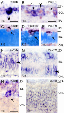
          All cadherins investigated by us are expressed differentially by restricted populations of retinal cells during specific periods of the ferret retinogenesis. For example, during embryonic development, some cadherins are exclusively expressed in the outer, proliferative zone of the neuroblast layer, whereas other cadherins mark the prospective ganglion cell layer or cells in the prospective inner nuclear layer. These expression patterns anticipate histogenetic changes that become visible in Nissl or nuclear stainings at later stages. In parallel to the ongoing development of retinal circuits, cadherin expression becomes restricted to specific subpopulations of retinal cell types, especially of ganglion cells, which express most of the investigated cadherins until adulthood. A comparison to previous results in chicken and mouse reveals overall conserved expression patterns of some cadherins but also species differences.

          Conclusions

          The spatiotemporally restricted expression patterns of 7 classic cadherins and 8 δ-protocadherins indicate that cadherins provide a combinatorial adhesive code that specifies developing retinal cell populations and intraretinal as well as retinofugal neural circuits in the developing ferret retina.

          Related collections

          Most cited references44

          • Record: found
          • Abstract: found
          • Article: not found

          Regulation of cadherin-mediated adhesion in morphogenesis.

          Cadherin cell-adhesion proteins mediate many facets of tissue morphogenesis. The dynamic regulation of cadherins in response to various extracellular signals controls cell sorting, cell rearrangements and cell movements. Cadherins are regulated at the cell surface by an inside-out signalling mechanism that is analogous to the integrins in platelets and leukocytes. Signal-transduction pathways impinge on the catenins (cytoplasmic cadherin-associated proteins), which transduce changes across the membrane to alter the state of the cadherin adhesive bond.
            Bookmark
            • Record: found
            • Abstract: found
            • Article: not found

            Cadherins in development: cell adhesion, sorting, and tissue morphogenesis.

            Tissue morphogenesis during development is dependent on activities of the cadherin family of cell-cell adhesion proteins that includes classical cadherins, protocadherins, and atypical cadherins (Fat, Dachsous, and Flamingo). The extracellular domain of cadherins contains characteristic repeats that regulate homophilic and heterophilic interactions during adhesion and cell sorting. Although cadherins may have originated to facilitate mechanical cell-cell adhesion, they have evolved to function in many other aspects of morphogenesis. These additional roles rely on cadherin interactions with a wide range of binding partners that modify their expression and adhesion activity by local regulation of the actin cytoskeleton and diverse signaling pathways. Here we examine how different members of the cadherin family act in different developmental contexts, and discuss the mechanisms involved.
              Bookmark
              • Record: found
              • Abstract: found
              • Article: not found

              Phylogenetic analysis of the cadherin superfamily allows identification of six major subfamilies besides several solitary members.

              Cadherins play an important role in specific cell-cell adhesion events. Their expression appears to be tightly regulated during development and each tissue or cell type shows a characteristic pattern of cadherin molecules. Inappropriate regulation of their expression levels or functionality has been observed in human malignancies, in many cases leading to aggravated cancer cell invasion and metastasis. The cadherins form a superfamily with at least six subfamilies, which can be distinguished on the basis of protein domain composition, genomic structure, and phylogenetic analysis of the protein sequences. These subfamilies comprise classical or type-I cadherins, atypical or type-II cadherins, desmocollins, desmogleins, protocadherins and Flamingo cadherins. In addition, several cadherins clearly occupy isolated positions in the cadherin superfamily (cadherin-13, -15, -16, -17, Dachsous, RET, FAT, MEGF1 and most invertebrate cadherins). We suggest a different evolutionary origin of the protocadherin and Flamingo cadherin genes versus the genes encoding desmogleins, desmocollins, classical cadherins, and atypical cadherins. The present phylogenetic analysis may accelerate the functional investigation of the whole cadherin superfamily by allowing focused research of prototype cadherins within each subfamily. Copyright 2000 Academic Press.
                Bookmark

                Author and article information

                Journal
                BMC Neurosci
                BMC Neuroscience
                BioMed Central
                1471-2202
                2009
                22 December 2009
                : 10
                : 153
                Affiliations
                [1 ]Institute of Anatomy I, University of Jena School of Medicine, Teichgraben 7, D-07743 Jena, Germany
                Article
                1471-2202-10-153
                10.1186/1471-2202-10-153
                2811116
                20028529
                d41b34aa-12d2-4dd2-9194-1472673c8605
                Copyright ©2009 Etzrodt et al; licensee BioMed Central Ltd.

                This is an Open Access article distributed under the terms of the Creative Commons Attribution License ( http://creativecommons.org/licenses/by/2.0), which permits unrestricted use, distribution, and reproduction in any medium, provided the original work is properly cited.

                History
                : 18 June 2009
                : 22 December 2009
                Categories
                Research article

                Neurosciences
                Neurosciences

                Comments

                Comment on this article