27
views
0
recommends
+1 Recommend
0 collections
    0
    shares
      • Record: found
      • Abstract: found
      • Article: found
      Is Open Access

      Immunohistochemistry for detection of avian infectious bronchitis virus strain M41 in the proventriculus and nervous system of experimentally infected chicken embryos

      research-article

      Read this article at

      Bookmark
          There is no author summary for this article yet. Authors can add summaries to their articles on ScienceOpen to make them more accessible to a non-specialist audience.

          Abstract

          Background

          Infectious bronchitis virus primarily induces a disease of the respiratory system, different IBV strains may show variable tissue tropisms and also affect the oviduct and the kidneys. Proventriculitis was also associated with some new IBV strains. Aim of this study was to investigate by immunohistochemistry (IHC) the tissue tropism of avian infectious bronchitis virus (IBV) strain M41 in experimentally infected chicken embryos.

          Results

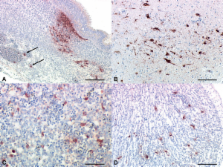
          To this end chicken embryos were inoculated in the allantoic sac with 10 3 EID 50 of IBV M41 at 10 days of age. At 48, 72, and 120 h postinoculation (PI), embryos and chorioallantoic membranes (CAM) were sampled, fixed, and paraffin-wax embedded. Allantoic fluid was also collected and titrated in chicken embryo kidney cells (CEK). The sensitivity of IHC in detecting IBV antigens in the CAM of inoculated eggs matched the virus reisolation and detection in CEK. Using IHC, antigens of IBV were detected in nasal epithelium, trachea, lung, spleen, myocardial vasculature, liver, gastrointestinal tract, kidney, skin, sclera of the eye, spinal cord, as well as in brain neurons of the inoculated embryos. These results were consistent with virus isolation and denote the wide tissue tropism of IBV M41 in the chicken embryo. Most importantly, we found infection of vasculature and smooth muscle of the proventriculus which has not seen before with IBV strain M41.

          Conclusion

          IHC can be an additional useful tool for diagnosis of IBV infection in chickens and allows further studies to foster a deeper understanding of the pathogenesis of infections with IBV strains of different virulence. Moreover, these results underline that embryonic tissues in addition to CAM could be also used as possible source to generate IBV antigens for diagnostic purposes.

          Related collections

          Most cited references45

          • Record: found
          • Abstract: found
          • Article: not found

          Coronaviruses in poultry and other birds.

          The number of avian species in which coronaviruses have been detected has doubled in the past couple of years. While the coronaviruses in these species have all been in coronavirus Group 3, as for the better known coronaviruses of the domestic fowl (infectious bronchitis virus [IBV], in Gallus gallus), turkey (Meleagris gallopavo) and pheasant (Phasianus colchicus), there is experimental evidence to suggest that birds are not limited to infection with Group 3 coronaviruses. In China coronaviruses have been isolated from peafowl (Pavo), guinea fowl (Numida meleagris; also isolated in Brazil), partridge (Alectoris) and also from a non-gallinaceous bird, the teal (Anas), all of which were being reared in the vicinity of domestic fowl. These viruses were closely related in genome organization and in gene sequences to IBV. Indeed, gene sequencing and experimental infection of chickens indicated that the peafowl isolate was the H120 IB vaccine strain, while the teal isolate was possibly a field strain of a nephropathogenic IBV. Thus the host range of IBV does extend beyond the chicken. Most recently, Group 3 coronaviruses have been detected in greylag goose (Anser anser), mallard duck (Anas platyrhynchos) and pigeon (Columbia livia). It is clear from the partial genome sequencing of these viruses that they are not IBV, as they have two additional small genes near the 3' end of the genome. Twenty years ago a coronavirus was isolated after inoculation of mice with tissue from the coastal shearwater (Puffinus puffinus). While it is not certain whether the virus was actually from the shearwater or from the mice, recent experiments have shown that bovine coronavirus (a Group 2 coronavirus) can infect and also cause enteric disease in turkeys. Experiments with some Group 1 coronaviruses (all from mammals, to date) have shown that they are not limited to replicating or causing disease in a single host. SARS-coronavirus has a wide host range. Clearly there is the potential for the emergence of new coronavirus diseases in domestic birds, from both avian and mammalian sources. Modest sequence conservation within gene 1 has enabled the design of oligonucleotide primers for use in diagnostic reverse transcriptase-polymerase chain reactions, which will be useful for the detection of new coronaviruses.
            Bookmark
            • Record: found
            • Abstract: found
            • Article: not found

            Recombinant avian infectious bronchitis virus expressing a heterologous spike gene demonstrates that the spike protein is a determinant of cell tropism.

            A recombinant infectious bronchitis virus (IBV), BeauR-M41(S), was generated using our reverse genetics system (R. Casais, V. Thiel, S. G. Siddell, D. Cavanagh, and P. Britton, J. Virol. 75:12359-12369, 2001), in which the ectodomain region of the spike gene from IBV M41-CK replaced the corresponding region of the IBV Beaudette genome. BeauR-M41(S) acquired the same cell tropism phenotype as IBV M41-CK in four different cell types, demonstrating that the IBV spike glycoprotein is a determinant of cell tropism.
              Bookmark
              • Record: found
              • Abstract: found
              • Article: not found

              A new genotype of nephropathogenic infectious bronchitis virus circulating in vaccinated and non-vaccinated flocks in China

              Five strains of infectious bronchitis virus (IBV) were isolated from five layer flocks that had nephropathogenic infection in four provinces in China. Among them, three of the five flocks had been vaccinated against infectious bronchitis. Virulence studies indicated that the five Chinese IBV isolates caused 10 to 30% mortality in 15-day-old specific pathogen free chickens and gross lesions were mainly confined to the kidneys in all of the dead chickens. Two oligonucleotide pairs, S1Uni2 and S1Oligo3′ or S1Oligo5′ and S1Oligo3′, were used after propagation of the isolates in embryonated eggs to amplify the S1 protein genes of the spike protein. The cDNA derived by reverse transcriptase-polymerase chain reaction was cloned and sequenced. The nucleotide and amino acid sequence of S1 protein gene had a similar degree of identity (≥92%) among the five Chinese IBV isolates. The nucleotide and amino acid identity of the S1 protein gene between the five Chinese IBV isolates and 16 strains of other IBVs varied from 60 to 81%. This clearly showed that the five Chinese IBV isolates comprised a separate genotype. These results demonstrated, for the first time, that there is a new genotype of nephropathogenic IBV circulating in vaccinated and non-vaccinated flocks in China.
                Bookmark

                Author and article information

                Journal
                Virol J
                Virology Journal
                BioMed Central
                1743-422X
                2009
                5 February 2009
                : 6
                : 15
                Affiliations
                [1 ]Virology Department, Faculty of Veterinary Medicine, Beni-Suef University, Beni-Suef 62511, Egypt
                [2 ]Setor de Patologia Veterinária, Faculdade de Veterinária – UFRGS, Av. Bento Gonçalves, 9090, Porto Alegre, RS, Brasil
                [3 ]Friedrich-Loeffler-Institut (FLI), Federal Research Institute for Animal Health, Südufer 10, 17493 Greifswald-Insel Riems, Germany
                Article
                1743-422X-6-15
                10.1186/1743-422X-6-15
                2657138
                19196466
                d0aa4c67-8403-4a79-83eb-a5512f830c15
                Copyright © 2009 Abdel-Moneim et al; licensee BioMed Central Ltd.

                This is an Open Access article distributed under the terms of the Creative Commons Attribution License ( http://creativecommons.org/licenses/by/2.0), which permits unrestricted use, distribution, and reproduction in any medium, provided the original work is properly cited.

                History
                : 1 December 2008
                : 5 February 2009
                Categories
                Research

                Microbiology & Virology
                Microbiology & Virology

                Comments

                Comment on this article