40
views
0
recommends
+1 Recommend
0 collections
    0
    shares
      • Record: found
      • Abstract: found
      • Article: found
      Is Open Access

      Onychomycosis Due to Nondermatophytic Molds

      research-article

      Read this article at

      Bookmark
          There is no author summary for this article yet. Authors can add summaries to their articles on ScienceOpen to make them more accessible to a non-specialist audience.

          Abstract

          Background

          Although there have been many studies about onychomycosis due to nondermatophytic molds (NDM), few studies about etiologic agents including NDM in onychomycosis have been reported in Korea. Objective: This study investigated onychomycosis due to NDM in the Gyeongju area of Korea.

          Objective

          This study investigated onychomycosis due to NDM in the Gyeongju area of Korea.

          Methods

          In the 10-year period from 1999~2009, we reviewed 59 patients with onychomycosis due to NDM. The etiologic agents were identified by cultures on Sabouraud's Dextrose agar with and without cycloheximide. In some cases, internal transcribed spacer sequence analysis was done. NDM isolated considered pathogens when the presence of fungal elements was identified by direct microscopy observation and in follow-up cultures yielding the same fungi.

          Results

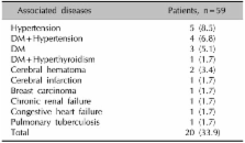
          Onychomycosis due to NDM comprised 2.3% of all onychomycosis. Of the 59 patients with onychomycosis due to NDM, 84.7% were toenail onychomycosis and 15.3% were fingernail onychomycosis. The incidence rate was highest in the fifth decade (27.1%). The ratio of male to female patients was 1:1.6. The frequency of associated diseases, in descending order, was hypertension, diabetes mellitus, and cerebral hematoma. Distal and lateral subungual onychomycosis (86.4%) was the most common clinical type of onychomycosis. Aspergillus spp. was the most frequently isolated etiologic agent of onychomycosis due to NDM (83.0%). Other causative agents were Scopulariopsis brevicaulis (10.2%), Acremonium spp. (3.4%), Fusarium solani (1.7%), and Chaetomium globosum (1.7%).

          Conclusion

          Because of the increase in onychomycosis due to NDM, we suggest the need of a careful mycological examination in patients with onychomycosis.

          Related collections

          Most cited references36

          • Record: found
          • Abstract: found
          • Article: not found

          Onychomycosis caused by nondermatophytic molds: clinical features and response to treatment of 59 cases.

          Nail invasion by nondermatophytic molds (NDM) is considered uncommon with prevalence rates ranging from 1.45% to 17. 6%. We report the clinical features and response to treatment of onychomycosis caused by these molds. From 1995 through 1998 we performed a mycologic study on 1548 patients affected by nail disorders, and we diagnosed 431 cases of onychomycosis including 59 cases of onychomycosis caused by molds. These include 17 patients with onychomycosis caused by Scopulariopsis brevicaulis, 26 patients with onychomycosis caused by Fusarium sp, 9 patients with onychomycosis caused by Acremonium sp, and 7 patients with onychomycosis caused by Aspergillus sp. Onychomycosis caused by S brevicaulis, Fusarium sp, and Aspergillus sp may often be suspected by clinical examination. In fact 38 of 50 patients with onychomycosis resulting from these molds were affected by proximal subungual onychomycosis associated with inflammation of the proximal nailfold. In our experience mold onychomycosis is not significantly associated with systemic diseases or immunodepression. NDM are difficult to eradicate; by using and combining different treatments (systemic itraconazole, systemic terbinafine, topical terbinafine after nail plate avulsion, and ciclopirox nail lacquer) we were able to cure only 69.2% of patients with S brevicaulis onychomycosis, 71.4% of patients with Acremonium onychomycosis, and 40% of patients with Fusarium onychomycosis. Aspergillus onychomycosis, on the other hand, responded very well to therapy and all our patients were cured after systemic or topical treatment. Eradication of the mold produced a complete cure of the nail abnormalities in all the patients who responded to treatment. Clinical examination usually suggests diagnosis of onychomycosis resulting from NDM. Topical treatment can be more successful than systemic therapy to cure onychomycosis caused by S brevicaulis, Fusarium sp, and Acremonium sp.
            Bookmark
            • Record: found
            • Abstract: found
            • Article: not found

            Other fungi causing onychomycosis.

            Nondermatophyte onychomycosis account for 2% to 12% of all nail fungal infections and can be caused by a wide range of fungi, mainly Scopulariopsis brevicaulis, Aspergillus versicolor, A. flavus, A. niger, A. fumigatus, Fusarium solani, F. oxysporum and Scytalidium spp. Among the predisposing factors are footwear, hyperhidrosis, local trauma, peripheral circulatory disease, and immunosuppression. These nondermatophyte fungi lack the keratinolytic capacity of dermatophytes, but they still can infect alone or in combination with the latter. Because most are considered laboratory contaminants, special criteria have been created for the correct diagnosis of nondermatophyte onychomycosis. The etiologic agent does not determine the clinical pattern of nail invasion, but superficial onychomycosis is frequently observed; leukonychia and melanonychia can also be clinical manifestations. Copyright 2010. Published by Elsevier Inc.
              Bookmark
              • Record: found
              • Abstract: found
              • Article: not found

              A U.S. epidemiologic survey of superficial fungal diseases.

              Large-scale studies performed outside the United States have demonstrated that most cases of onychomycosis and tinea pedis are caused by dermatophytes, primarily Trichophyton rubrum. However, other studies have suggested that yeasts and nondermatophytic molds may play a role, particularly in onychomycosis. This study was undertaken to determine the epidemiology of superficial fungal infections in a U.S. population. Fungal cultures were performed on patients with clinically suspected tinea cruris, tinea corporis, tinea capitis, tinea pedis, and onychomycosis. Dermatophytes were the most commonly isolated fungi in each type of superficial fungal disease studied. T. rubrum was the most commonly isolated dermatophyte species, although Trichophyton tonsurans was more common in tinea capitis and equally common in tinea corporis/tinea cruris. In tinea pedis and onychomycosis, dermatophytes appeared in approximately 95% and 82% of isolates, respectively. Candida albicans and nondermatophyte molds played only a minor role in onychomycosis; C. albicans was isolated in 7% of nail cultures and nondermatophytic molds were isolated in 11%. These results are in general agreement with other major epidemiologic studies performed outside the United States. Dermatophyte fungi cause most superficial fungal infections.
                Bookmark

                Author and article information

                Journal
                Ann Dermatol
                Ann Dermatol
                AD
                Annals of Dermatology
                Korean Dermatological Association; The Korean Society for Investigative Dermatology
                1013-9087
                2005-3894
                May 2012
                26 April 2012
                : 24
                : 2
                : 175-180
                Affiliations
                Department of Dermatology, Dongguk University College of Medicine, Gyeongju, Korea.
                [1 ]Department of Laboratory Medicine, Dongguk University College of Medicine, Gyeongju, Korea.
                Author notes
                Corresponding author: Moo Kyu Suh, M.D., Department of Dermatology, Gyeongju Hospital, College of Medicine, Dongguk University, 707 Seokjang-dong, Gyeongju 780-350, Korea. Tel: 82-54-770-8268, Fax: 82-54-773-1581, smg@ 123456dongguk.ac.kr
                Article
                10.5021/ad.2012.24.2.175
                3346908
                22577268
                ca384ba0-03de-4cdf-ba72-7de7e398e465
                Copyright © 2012 Korean Dermatological Association; The Korean Society for Investigative Dermatology

                This is an Open Access article distributed under the terms of the Creative Commons Attribution Non-Commercial License ( http://creativecommons.org/licenses/by-nc/3.0) which permits unrestricted non-commercial use, distribution, and reproduction in any medium, provided the original work is properly cited.

                History
                : 13 May 2011
                : 19 July 2011
                : 12 August 2011
                Categories
                Original Article

                Dermatology
                onychomycosis,molds
                Dermatology
                onychomycosis, molds

                Comments

                Comment on this article