7
views
0
recommends
+1 Recommend
0 collections
    0
    shares
      • Record: found
      • Abstract: found
      • Article: found
      Is Open Access

      Taphonomic patterns mimic biologic structures: diagenetic Liesegang rings in Mesozoic coleoids and coprolites

      research-article

      Read this article at

      Bookmark
          There is no author summary for this article yet. Authors can add summaries to their articles on ScienceOpen to make them more accessible to a non-specialist audience.

          Abstract

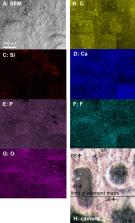
          Because of physiology of coleoids, their fossils preserve soft-tissue-remains more often than other cephalopods. Sometimes, the phosphatized soft-tissues, particularly parts of the muscular mantle, display dark circular patterns. Here, we showcase that these patterns, here documented for fossil coleoids from the Jurassic of Germany and the Cretaceous of Lebanon, superficially resemble chromatophores (which enable living coleoids to alter their coloration). We examined and chemically analyzed the circular structures in these specimens, describe them, and discuss their genesis. Based on their structure and color, we visually differentiate between three types of circles. By comparison with similar structures, we suggest that these structures are not biogenic but Liesegang rings, which formed due to reaction-diffusion processes very soon after death.

          Related collections

          Most cited references63

          • Record: found
          • Abstract: found
          • Article: not found

          Direct chemical evidence for eumelanin pigment from the Jurassic period.

          Melanin is a ubiquitous biological pigment found in bacteria, fungi, plants, and animals. It has a diverse range of ecological and biochemical functions, including display, evasion, photoprotection, detoxification, and metal scavenging. To date, evidence of melanin in fossil organisms has relied entirely on indirect morphological and chemical analyses. Here, we apply direct chemical techniques to categorically demonstrate the preservation of eumelanin in two > 160 Ma Jurassic cephalopod ink sacs and to confirm its chemical similarity to the ink of the modern cephalopod, Sepia officinalis. Identification and characterization of degradation-resistant melanin may provide insights into its diverse roles in ancient organisms.
            Bookmark
            • Record: found
            • Abstract: not found
            • Article: not found

            The role of the calcium carbonate-calcium phosphate switch in the mineralization of soft-bodied fossils

              Bookmark
              • Record: found
              • Abstract: found
              • Article: not found

              Advanced chemical methods in melanin determination.

              Among the biopolymers, melanins are unique in many respects. The other essential biopolymers - proteins, nucleic acids, and carbohydrates - are chemically well characterized; their precursors (monomer units) and modes of connection between the monomer units are known, and sequences of their connection can be determined with well-established methodologies. In contrast, we still do not have a method to determine accurately the ratio of various units present in melanins. This is largely because of the chemical properties of melanins, such as their insolubility over a broad range of pH, the heterogeneity in their structural features, and also because of the lack of methods that can split melanin polymers into their monomer units (all other biopolymers can be hydrolysed to the corresponding monomer units). To overcome this difficulty, we developed a rapid and sensitive method for quantitatively analysing eumelanin and pheomelanin in biological samples by chemical degradation methods followed by HPLC determination. This HPLC microanalytical method for characterizing eumelanin and pheomelanin has become a useful tool for the study of melanogenesis. This review will summarize the usefulness and limitations of the various chemical and spectrophotometric methods used to analyse melanins at the biochemical, cellular, and tissue levels. Emphasis is given on the usefulness of 4-amino-3-hydroxyphenylalanine as a specific marker of pheomelanin.
                Bookmark

                Author and article information

                Contributors
                Journal
                PeerJ
                PeerJ
                PeerJ
                PeerJ
                PeerJ
                PeerJ Inc. (San Diego, USA )
                2167-8359
                14 January 2021
                2021
                : 9
                : e10703
                Affiliations
                [1 ]Paläontologisches Institut und Museum, Universität Zürich , Zürich, Switzerland
                [2 ]Trilobite Design Italia , Aurisina (TS), Italy
                [3 ]Institute of Geology, Mineralogy & Geophysics, Ruhr-Universität Bochum , Bochum, Germany
                [4 ]Staatliches Museum für Naturkunde , Stuttgart, Germany
                [5 ]Bayerische Staatssammlung für Paläontologie und Geologie , München, Germany
                [6 ]School of Geography, Earth and Environmental Sciences, University of Birmingham , Birmingham, UK
                [7 ]Institute of Earth Sciences (ISTE), University of Lausanne , Lausanne, Switzerland
                Author information
                http://orcid.org/0000-0002-6563-4720
                http://orcid.org/0000-0002-7529-3456
                Article
                10703
                10.7717/peerj.10703
                7811783
                33520466
                bfc71814-143e-4545-a41e-189e7ceebd39
                © 2021 Klug et al.

                This is an open access article distributed under the terms of the Creative Commons Attribution License, which permits unrestricted use, distribution, reproduction and adaptation in any medium and for any purpose provided that it is properly attributed. For attribution, the original author(s), title, publication source (PeerJ) and either DOI or URL of the article must be cited.

                History
                : 20 August 2020
                : 14 December 2020
                Funding
                Funded by: Swiss National Science Foundation SNF
                Award ID: 200021_169627
                This research was supported by the Swiss National Science Foundation SNF (grant 200021_169627). The funders had no role in study design, data collection and analysis, decision to publish, or preparation of the manuscript.
                Categories
                Marine Biology
                Paleontology

                coleoidea,vampyropoda,jurassic,cretaceous,diagenesis,exceptional preservation,conservation deposits

                Comments

                Comment on this article