76
views
0
recommends
+1 Recommend
0 collections
    0
    shares
      • Record: found
      • Abstract: found
      • Article: not found

      Simultaneous all-optical manipulation and recording of neural circuit activity with cellular resolution in vivo

      research-article
      , , ,
      Nature methods

      Read this article at

      ScienceOpenPublisherPMC
      Bookmark
          There is no author summary for this article yet. Authors can add summaries to their articles on ScienceOpen to make them more accessible to a non-specialist audience.

          Abstract

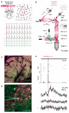
          We describe an all-optical strategy for simultaneously manipulating and recording the activity of multiple neurons with cellular resolution in vivo. Concurrent two-photon optogenetic activation and calcium imaging is enabled by coexpression of a red-shifted opsin and a genetically encoded calcium indicator. A spatial light modulator allows tens of user-selected neurons to be targeted for spatiotemporally precise optogenetic activation, while simultaneous fast calcium imaging provides high-resolution network-wide readout of the manipulation with negligible optical crosstalk. Proof-of-principle experiments in mouse barrel cortex demonstrate interrogation of the same neuronal population during different behavioral states, and targeting of neuronal ensembles based on their functional signature. This approach extends the optogenetic toolkit beyond the specificity obtained with genetic or viral approaches, enabling high-throughput, flexible and long-term optical interrogation of functionally defined neural circuits with single-cell and single-spike resolution in the mammalian brain in vivo.

          Related collections

          Most cited references37

          • Record: found
          • Abstract: found
          • Article: not found

          Neocortical excitation/inhibition balance in information processing and social dysfunction.

          Severe behavioural deficits in psychiatric diseases such as autism and schizophrenia have been hypothesized to arise from elevations in the cellular balance of excitation and inhibition (E/I balance) within neural microcircuitry. This hypothesis could unify diverse streams of pathophysiological and genetic evidence, but has not been susceptible to direct testing. Here we design and use several novel optogenetic tools to causally investigate the cellular E/I balance hypothesis in freely moving mammals, and explore the associated circuit physiology. Elevation, but not reduction, of cellular E/I balance within the mouse medial prefrontal cortex was found to elicit a profound impairment in cellular information processing, associated with specific behavioural impairments and increased high-frequency power in the 30-80 Hz range, which have both been observed in clinical conditions in humans. Consistent with the E/I balance hypothesis, compensatory elevation of inhibitory cell excitability partially rescued social deficits caused by E/I balance elevation. These results provide support for the elevated cellular E/I balance hypothesis of severe neuropsychiatric disease-related symptoms.
            Bookmark
            • Record: found
            • Abstract: not found
            • Article: not found

            Two-photon laser scanning fluorescence microscopy

              Bookmark
              • Record: found
              • Abstract: found
              • Article: not found

              Optogenetic stimulation of a hippocampal engram activates fear memory recall

              A specific memory is thought to be encoded by a sparse population of neurons 1,2 . These neurons can be tagged during learning for subsequent identification 3 and manipulation 4,5,6 . Moreover, their ablation or inactivation results in reduced memory expression, suggesting their necessity in mnemonic processes. However, a critical question of sufficiency remains: can one elicit the behavioral output of a specific memory by directly activating a population of neurons that was active during learning? Here we show that optogenetic reactivation of hippocampal neurons activated during fear conditioning is sufficient to induce freezing behavior. We labeled a population of hippocampal dentate gyrus neurons activated during fear learning with channelrhodopsin-2 (ChR2) 7,8 and later optically reactivated these neurons in a different context. The mice showed increased freezing only upon light stimulation, indicating light-induced fear memory recall. This freezing was not detected in non-fear conditioned mice expressing ChR2 in a similar proportion of cells, nor in fear conditioned mice with cells labeled by EYFP instead of ChR2. Finally, activation of cells labeled in a context not associated with fear did not evoke freezing in mice that were previously fear conditioned in a different context, suggesting that light-induced fear memory recall is context-specific. Together, our findings indicate that activating a sparse but specific ensemble of hippocampal neurons that contribute to a memory engram is sufficient for the recall of that memory. Moreover, our experimental approach offers a general method of mapping cellular populations bearing memory engrams.
                Bookmark

                Author and article information

                Contributors
                Journal
                101215604
                32338
                Nat Methods
                Nat. Methods
                Nature methods
                1548-7091
                1548-7105
                29 June 2016
                22 December 2014
                February 2015
                05 July 2016
                : 12
                : 2
                : 140-146
                Affiliations
                Wolfson Institute for Biomedical Research, University College London, Gower Street, London WC1E 6BT, UK; Department of Neuroscience, Physiology and Pharmacology, University College London, Gower Street, London WC1E 6BT, UK
                Wolfson Institute for Biomedical Research, University College London, Gower Street, London WC1E 6BT, UK; Department of Neuroscience, Physiology and Pharmacology, University College London, Gower Street, London WC1E 6BT, UK
                Wolfson Institute for Biomedical Research, University College London, Gower Street, London WC1E 6BT, UK; Department of Neuroscience, Physiology and Pharmacology, University College London, Gower Street, London WC1E 6BT, UK
                Wolfson Institute for Biomedical Research, University College London, Gower Street, London WC1E 6BT, UK; Department of Neuroscience, Physiology and Pharmacology, University College London, Gower Street, London WC1E 6BT, UK
                Author notes
                Address for correspondence: Michael Häusser: m.hausser@ 123456ucl.ac.uk , tel. +44-(0)20-7679-6756
                Article
                EMS61225
                10.1038/nmeth.3217
                4933203
                25532138
                bc044b8b-3a41-4f26-ad4d-bb0c6798850a

                Users may view, print, copy, and download text and data-mine the content in such documents, for the purposes of academic research, subject always to the full Conditions of use: http://www.nature.com/authors/editorial_policies/license.html#terms

                History
                Categories
                Article

                Life sciences
                Life sciences

                Comments

                Comment on this article