277
views
0
recommends
+1 Recommend
1 collections
    0
    shares
      • Record: found
      • Abstract: found
      • Article: found
      Is Open Access

      Structural basis for the recognition of SARS-CoV-2 by full-length human ACE2

      research-article

      Read this article at

      Bookmark
          There is no author summary for this article yet. Authors can add summaries to their articles on ScienceOpen to make them more accessible to a non-specialist audience.

          How SARS-CoV-2 binds to human cells

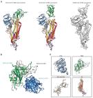
          Scientists are racing to learn the secrets of severe acute respiratory syndrome–coronavirus 2 (SARS-CoV-2), which is the cause of the pandemic disease COVID-19. The first step in viral entry is the binding of the viral trimeric spike protein to the human receptor angiotensin-converting enzyme 2 (ACE2). Yan et al. present the structure of human ACE2 in complex with a membrane protein that it chaperones, B 0AT1. In the context of this complex, ACE2 is a dimer. A further structure shows how the receptor binding domain of SARS-CoV-2 interacts with ACE2 and suggests that it is possible that two trimeric spike proteins bind to an ACE2 dimer. The structures provide a basis for the development of therapeutics targeting this crucial interaction.

          Science, this issue p. 1444

          Abstract

          Insight into how SARS-CoV-2 binds its human receptor could provide a basis for the development of therapeutics.

          Abstract

          Angiotensin-converting enzyme 2 (ACE2) is the cellular receptor for severe acute respiratory syndrome–coronavirus (SARS-CoV) and the new coronavirus (SARS-CoV-2) that is causing the serious coronavirus disease 2019 (COVID-19) epidemic. Here, we present cryo–electron microscopy structures of full-length human ACE2 in the presence of the neutral amino acid transporter B 0AT1 with or without the receptor binding domain (RBD) of the surface spike glycoprotein (S protein) of SARS-CoV-2, both at an overall resolution of 2.9 angstroms, with a local resolution of 3.5 angstroms at the ACE2-RBD interface. The ACE2-B 0AT1 complex is assembled as a dimer of heterodimers, with the collectrin-like domain of ACE2 mediating homodimerization. The RBD is recognized by the extracellular peptidase domain of ACE2 mainly through polar residues. These findings provide important insights into the molecular basis for coronavirus recognition and infection.

          Related collections

          Most cited references16

          • Record: found
          • Abstract: found
          • Article: not found

          A Novel Coronavirus from Patients with Pneumonia in China, 2019

          Summary In December 2019, a cluster of patients with pneumonia of unknown cause was linked to a seafood wholesale market in Wuhan, China. A previously unknown betacoronavirus was discovered through the use of unbiased sequencing in samples from patients with pneumonia. Human airway epithelial cells were used to isolate a novel coronavirus, named 2019-nCoV, which formed a clade within the subgenus sarbecovirus, Orthocoronavirinae subfamily. Different from both MERS-CoV and SARS-CoV, 2019-nCoV is the seventh member of the family of coronaviruses that infect humans. Enhanced surveillance and further investigation are ongoing. (Funded by the National Key Research and Development Program of China and the National Major Project for Control and Prevention of Infectious Disease in China.)
            Bookmark
            • Record: found
            • Abstract: found
            • Article: found
            Is Open Access

            A pneumonia outbreak associated with a new coronavirus of probable bat origin

            Since the outbreak of severe acute respiratory syndrome (SARS) 18 years ago, a large number of SARS-related coronaviruses (SARSr-CoVs) have been discovered in their natural reservoir host, bats 1–4 . Previous studies have shown that some bat SARSr-CoVs have the potential to infect humans 5–7 . Here we report the identification and characterization of a new coronavirus (2019-nCoV), which caused an epidemic of acute respiratory syndrome in humans in Wuhan, China. The epidemic, which started on 12 December 2019, had caused 2,794 laboratory-confirmed infections including 80 deaths by 26 January 2020. Full-length genome sequences were obtained from five patients at an early stage of the outbreak. The sequences are almost identical and share 79.6% sequence identity to SARS-CoV. Furthermore, we show that 2019-nCoV is 96% identical at the whole-genome level to a bat coronavirus. Pairwise protein sequence analysis of seven conserved non-structural proteins domains show that this virus belongs to the species of SARSr-CoV. In addition, 2019-nCoV virus isolated from the bronchoalveolar lavage fluid of a critically ill patient could be neutralized by sera from several patients. Notably, we confirmed that 2019-nCoV uses the same cell entry receptor—angiotensin converting enzyme II (ACE2)—as SARS-CoV.
              Bookmark
              • Record: found
              • Abstract: found
              • Article: found
              Is Open Access

              Cryo-EM structure of the 2019-nCoV spike in the prefusion conformation

              Structure of the nCoV trimeric spike The World Health Organization has declared the outbreak of a novel coronavirus (2019-nCoV) to be a public health emergency of international concern. The virus binds to host cells through its trimeric spike glycoprotein, making this protein a key target for potential therapies and diagnostics. Wrapp et al. determined a 3.5-angstrom-resolution structure of the 2019-nCoV trimeric spike protein by cryo–electron microscopy. Using biophysical assays, the authors show that this protein binds at least 10 times more tightly than the corresponding spike protein of severe acute respiratory syndrome (SARS)–CoV to their common host cell receptor. They also tested three antibodies known to bind to the SARS-CoV spike protein but did not detect binding to the 2019-nCoV spike protein. These studies provide valuable information to guide the development of medical counter-measures for 2019-nCoV. Science, this issue p. 1260
                Bookmark

                Author and article information

                Journal
                Science
                Science
                SCIENCE
                science
                Science (New York, N.y.)
                American Association for the Advancement of Science
                0036-8075
                1095-9203
                27 March 2020
                04 March 2020
                : 367
                : 6485
                : 1444-1448
                Affiliations
                [1 ]Key Laboratory of Structural Biology of Zhejiang Province, Institute of Biology, Westlake Institute for Advanced Study, 18 Shilongshan Road, Hangzhou 310024, Zhejiang Province, China.
                [2 ]School of Life Sciences, Westlake University, 18 Shilongshan Road, Hangzhou 310024, Zhejiang Province, China.
                [3 ]Beijing Advanced Innovation Center for Structural Biology, Tsinghua-Peking Joint Center for Life Sciences, School of Life Sciences, Tsinghua University, Beijing 100084, China.
                Author notes
                [*]

                These authors contributed equally to this work.

                []Corresponding author. Email: zhouqiang@ 123456westlake.edu.cn
                Author information
                https://orcid.org/0000-0002-7122-6547
                https://orcid.org/0000-0002-2500-0010
                https://orcid.org/0000-0003-3007-3086
                https://orcid.org/0000-0002-6237-8813
                Article
                abb2762
                10.1126/science.abb2762
                7164635
                32132184
                bbd4e804-8f23-4ebb-aa1a-f51f1b11d4ff
                Copyright © 2020 The Authors, some rights reserved; exclusive licensee American Association for the Advancement of Science. No claim to original U.S. Government Works

                This open access article is distributed under Creative Commons Attribution License 4.0 (CC BY).

                History
                : 12 February 2020
                : 03 March 2020
                Funding
                Funded by: doi http://dx.doi.org/10.13039/501100001809, National Natural Science Foundation of China;
                Award ID: 31971123
                Funded by: doi http://dx.doi.org/10.13039/501100001809, National Natural Science Foundation of China;
                Award ID: 81920108015
                Funded by: doi http://dx.doi.org/10.13039/501100001809, National Natural Science Foundation of China;
                Award ID: 31930059
                Funded by: Key R&D Program of Zhejiang Province;
                Award ID: 2020C04001
                Funded by: the SARS-CoV-2 emergency project of the Science and Technology Department of Zhejiang Province;
                Award ID: 2020C03129
                Categories
                Research Article
                Research Articles
                R-Articles
                Biochem
                Cell Biol
                Custom metadata
                5
                5
                Valda Vinson
                Amelia Beyna

                Uncategorized
                Uncategorized

                Comments

                Comment on this article