92
views
0
recommends
+1 Recommend
0 collections
    0
    shares
      • Record: found
      • Abstract: found
      • Article: found
      Is Open Access

      Regional gray matter volumetric changes in autism associated with social and repetitive behavior symptoms

      research-article

      Read this article at

      Bookmark
          There is no author summary for this article yet. Authors can add summaries to their articles on ScienceOpen to make them more accessible to a non-specialist audience.

          Abstract

          Background

          Although differences in brain anatomy in autism have been difficult to replicate using manual tracing methods, automated whole brain analyses have begun to find consistent differences in regions of the brain associated with the social cognitive processes that are often impaired in autism. We attempted to replicate these whole brain studies and to correlate regional volume changes with several autism symptom measures.

          Methods

          We performed MRI scans on 24 individuals diagnosed with DSM-IV autistic disorder and compared those to scans from 23 healthy comparison subjects matched on age. All participants were male. Whole brain, voxel-wise analyses of regional gray matter volume were conducted using voxel-based morphometry (VBM).

          Results

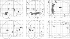
          Controlling for age and total gray matter volume, the volumes of the medial frontal gyri, left pre-central gyrus, right post-central gyrus, right fusiform gyrus, caudate nuclei and the left hippocampus were larger in the autism group relative to controls. Regions exhibiting smaller volumes in the autism group were observed exclusively in the cerebellum. Significant partial correlations were found between the volumes of the caudate nuclei, multiple frontal and temporal regions, the cerebellum and a measure of repetitive behaviors, controlling for total gray matter volume. Social and communication deficits in autism were also associated with caudate, cerebellar, and precuneus volumes, as well as with frontal and temporal lobe regional volumes.

          Conclusion

          Gray matter enlargement was observed in areas that have been functionally identified as important in social-cognitive processes, such as the medial frontal gyri, sensorimotor cortex and middle temporal gyrus. Additionally, we have shown that VBM is sensitive to associations between social and repetitive behaviors and regional brain volumes in autism.

          Related collections

          Most cited references80

          • Record: found
          • Abstract: not found
          • Article: not found

          Diagnostic and statistical manual of mental disorders.

            Bookmark
            • Record: found
            • Abstract: found
            • Article: not found

            A unified statistical approach for determining significant signals in images of cerebral activation.

            We present a unified statistical theory for assessing the significance of apparent signal observed in noisy difference images. The results are usable in a wide range of applications, including fMRI, but are discussed with particular reference to PET images which represent changes in cerebral blood flow elicited by a specific cognitive or sensorimotor task. Our main result is an estimate of the P-value for local maxima of Gaussian, t, chi(2) and F fields over search regions of any shape or size in any number of dimensions. This unifies the P-values for large search areas in 2-D (Friston et al. [1991]: J Cereb Blood Flow Metab 11:690-699) large search regions in 3-D (Worsley et al. [1992]: J Cereb Blood Flow Metab 12:900-918) and the usual uncorrected P-value at a single pixel or voxel. Copyright (c) 1996 Wiley-Liss, Inc.
              Bookmark
              • Record: found
              • Abstract: found
              • Article: not found

              Unusual brain growth patterns in early life in patients with autistic disorder: an MRI study.

              To quantify developmental abnormalities in cerebral and cerebellar volume in autism. The authors studied 60 autistic and 52 normal boys (age, 2 to 16 years) using MRI. Thirty autistic boys were diagnosed and scanned when 5 years or older. The other 30 were scanned when 2 through 4 years of age and then diagnosed with autism at least 2.5 years later, at an age when the diagnosis of autism is more reliable. Neonatal head circumferences from clinical records were available for 14 of 15 autistic 2- to 5-year-olds and, on average, were normal (35.1 +/- 1.3 cm versus clinical norms: 34.6 +/- 1.6 cm), indicative of normal overall brain volume at birth; one measure was above the 95th percentile. By ages 2 to 4 years, 90% of autistic boys had a brain volume larger than normal average, and 37% met criteria for developmental macrencephaly. Autistic 2- to 3-year-olds had more cerebral (18%) and cerebellar (39%) white matter, and more cerebral cortical gray matter (12%) than normal, whereas older autistic children and adolescents did not have such enlarged gray and white matter volumes. In the cerebellum, autistic boys had less gray matter, smaller ratio of gray to white matter, and smaller vermis lobules VI-VII than normal controls. Abnormal regulation of brain growth in autism results in early overgrowth followed by abnormally slowed growth. Hyperplasia was present in cerebral gray matter and cerebral and cerebellar white matter in early life in patients with autism.
                Bookmark

                Author and article information

                Journal
                BMC Psychiatry
                BMC Psychiatry
                BioMed Central (London )
                1471-244X
                2006
                13 December 2006
                : 6
                : 56
                Affiliations
                [1 ]Department of Psychiatry, University of Colorado Health Sciences Center, Denver, CO, 80220, USA
                [2 ]Department of Psychology, Colorado State University, Fort Collins, CO, 80523, USA
                [3 ]Department of Psychiatry and M.I.N.D. Institute, University of California at Davis, Sacramento, CA, 95817, USA
                Article
                1471-244X-6-56
                10.1186/1471-244X-6-56
                1770914
                17166273
                a7e9020b-470f-4929-8e1a-aba612a61f4a
                Copyright © 2006 Rojas et al; licensee BioMed Central Ltd.

                This is an Open Access article distributed under the terms of the Creative Commons Attribution License ( http://creativecommons.org/licenses/by/2.0), which permits unrestricted use, distribution, and reproduction in any medium, provided the original work is properly cited.

                History
                : 16 May 2006
                : 13 December 2006
                Categories
                Research Article

                Clinical Psychology & Psychiatry
                Clinical Psychology & Psychiatry

                Comments

                Comment on this article