14
views
0
recommends
+1 Recommend
1 collections
    0
    shares
      • Record: found
      • Abstract: found
      • Article: not found

      Deep metric learning-based image retrieval system for chest radiograph and its clinical applications in COVID-19

      research-article

      Read this article at

      ScienceOpenPublisherPMC
      Bookmark
          There is no author summary for this article yet. Authors can add summaries to their articles on ScienceOpen to make them more accessible to a non-specialist audience.

          Abstract

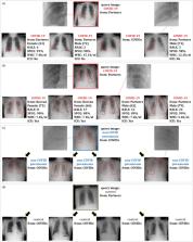
          In recent years, deep learning-based image analysis methods have been widely applied in computer-aided detection, diagnosis and prognosis, and has shown its value during the public health crisis of the novel coronavirus disease 2019 (COVID-19) pandemic. Chest radiograph (CXR) has been playing a crucial role in COVID-19 patient triaging, diagnosing and monitoring, particularly in the United States. Considering the mixed and unspecific signals in CXR, an image retrieval model of CXR that provides both similar images and associated clinical information can be more clinically meaningful than a direct image diagnostic model. In this work we develop a novel CXR image retrieval model based on deep metric learning. Unlike traditional diagnostic models which aim at learning the direct mapping from images to labels, the proposed model aims at learning the optimized embedding space of images, where images with the same labels and similar contents are pulled together. The proposed model utilizes multi-similarity loss with hard-mining sampling strategy and attention mechanism to learn the optimized embedding space, and provides similar images, the visualizations of disease-related attention maps and useful clinical information to assist clinical decisions. The model is trained and validated on an international multi-site COVID-19 dataset collected from 3 different sources. Experimental results of COVID-19 image retrieval and diagnosis tasks show that the proposed model can serve as a robust solution for CXR analysis and patient management for COVID-19. The model is also tested on its transferability on a different clinical decision support task for COVID-19, where the pre-trained model is applied to extract image features from a new dataset without any further training. The extracted features are then combined with COVID-19 patient's vitals, lab tests and medical histories to predict the possibility of airway intubation in 72 hours, which is strongly associated with patient prognosis, and is crucial for patient care and hospital resource planning. These results demonstrate our deep metric learning based image retrieval model is highly efficient in the CXR retrieval, diagnosis and prognosis, and thus has great clinical value for the treatment and management of COVID-19 patients.

          Graphical abstract

          Related collections

          Most cited references60

          • Record: found
          • Abstract: found
          • Article: not found

          A survey on deep learning in medical image analysis

          Deep learning algorithms, in particular convolutional networks, have rapidly become a methodology of choice for analyzing medical images. This paper reviews the major deep learning concepts pertinent to medical image analysis and summarizes over 300 contributions to the field, most of which appeared in the last year. We survey the use of deep learning for image classification, object detection, segmentation, registration, and other tasks. Concise overviews are provided of studies per application area: neuro, retinal, pulmonary, digital pathology, breast, cardiac, abdominal, musculoskeletal. We end with a summary of the current state-of-the-art, a critical discussion of open challenges and directions for future research.
            Bookmark
            • Record: found
            • Abstract: found
            • Article: not found

            Covid-19: automatic detection from X-ray images utilizing transfer learning with convolutional neural networks

            In this study, a dataset of X-ray images from patients with common bacterial pneumonia, confirmed Covid-19 disease, and normal incidents, was utilized for the automatic detection of the Coronavirus disease. The aim of the study is to evaluate the performance of state-of-the-art convolutional neural network architectures proposed over the recent years for medical image classification. Specifically, the procedure called Transfer Learning was adopted. With transfer learning, the detection of various abnormalities in small medical image datasets is an achievable target, often yielding remarkable results. The datasets utilized in this experiment are two. Firstly, a collection of 1427 X-ray images including 224 images with confirmed Covid-19 disease, 700 images with confirmed common bacterial pneumonia, and 504 images of normal conditions. Secondly, a dataset including 224 images with confirmed Covid-19 disease, 714 images with confirmed bacterial and viral pneumonia, and 504 images of normal conditions. The data was collected from the available X-ray images on public medical repositories. The results suggest that Deep Learning with X-ray imaging may extract significant biomarkers related to the Covid-19 disease, while the best accuracy, sensitivity, and specificity obtained is 96.78%, 98.66%, and 96.46% respectively. Since by now, all diagnostic tests show failure rates such as to raise concerns, the probability of incorporating X-rays into the diagnosis of the disease could be assessed by the medical community, based on the findings, while more research to evaluate the X-ray approach from different aspects may be conducted.
              Bookmark
              • Record: found
              • Abstract: found
              • Article: not found

              Squeeze-and-Excitation Networks

              The central building block of convolutional neural networks (CNNs) is the convolution operator, which enables networks to construct informative features by fusing both spatial and channel-wise information within local receptive fields at each layer. A broad range of prior research has investigated the spatial component of this relationship, seeking to strengthen the representational power of a CNN by enhancing the quality of spatial encodings throughout its feature hierarchy. In this work, we focus instead on the channel relationship and propose a novel architectural unit, which we term the "Squeeze-and-Excitation" (SE) block, that adaptively recalibrates channel-wise feature responses by explicitly modelling interdependencies between channels. We show that these blocks can be stacked together to form SENet architectures that generalise extremely effectively across different datasets. We further demonstrate that SE blocks bring significant improvements in performance for existing state-of-the-art CNNs at minimal additional computational cost. Squeeze-and-Excitation Networks formed the foundation of our ILSVRC 2017 classification submission which won first place and reduced the top-5 error to 2.251%, surpassing the winning entry of 2016 by a relative improvement of ∼25%. Models and code are available at https://github.com/hujie-frank/SENet.
                Bookmark

                Author and article information

                Journal
                Med Image Anal
                Med Image Anal
                Medical Image Analysis
                Published by Elsevier B.V.
                1361-8415
                1361-8423
                7 February 2021
                May 2021
                7 February 2021
                : 70
                : 101993
                Affiliations
                [a ]Department of Radiology, Massachusetts General Hospital, Boston, MA, United States
                [b ]School of Engineering and Applied Sciences, Harvard University, Boston, MA, United States
                [c ]MGH & BWH Center for Clinical Data Science, Boston, MA, United States
                [d ]Department of Internal Medicine, School of Medicine, Kyungpook National University, Daegu, South Korea
                [e ]Department of Internal Medicine, Yeungnam University College of Medicine, Daegu, South Korea
                [f ]Department of Internal Medicine, Catholic University of Daegu School of Medicine, Daegu, South Korea
                [g ]Department of Internal Medicine, Keimyung University School of Medicine, Daegu, South Korea
                Author notes
                [* ]Corresponding author.
                [1]

                These authors contribute equally to this work

                Article
                S1361-8415(21)00039-6 101993
                10.1016/j.media.2021.101993
                8032481
                33711739
                9a3aa690-354b-41ab-9011-cd7cb7acfa8b
                © 2021 Published by Elsevier B.V.

                Since January 2020 Elsevier has created a COVID-19 resource centre with free information in English and Mandarin on the novel coronavirus COVID-19. The COVID-19 resource centre is hosted on Elsevier Connect, the company's public news and information website. Elsevier hereby grants permission to make all its COVID-19-related research that is available on the COVID-19 resource centre - including this research content - immediately available in PubMed Central and other publicly funded repositories, such as the WHO COVID database with rights for unrestricted research re-use and analyses in any form or by any means with acknowledgement of the original source. These permissions are granted for free by Elsevier for as long as the COVID-19 resource centre remains active.

                History
                : 9 June 2020
                : 19 January 2021
                : 1 February 2021
                Categories
                Article

                Radiology & Imaging
                chest radiograph,covid-19,image retrieval,image content query
                Radiology & Imaging
                chest radiograph, covid-19, image retrieval, image content query

                Comments

                Comment on this article