2
views
0
recommends
+1 Recommend
0 collections
    0
    shares
      • Record: found
      • Abstract: found
      • Article: found
      Is Open Access

      The Role of the Negative Regulation of Microglia-Mediated Neuroinflammation in Improving Emotional Behavior After Epileptic Seizures

      research-article

      Read this article at

      Bookmark
          There is no author summary for this article yet. Authors can add summaries to their articles on ScienceOpen to make them more accessible to a non-specialist audience.

          Abstract

          Objective

          Studies have long shown that uncontrolled inflammatory responses in the brain play a key role in epilepsy pathogenesis. Microglias play an important role in epileptic-induced neuroinflammation, but their role after epileptic seizures is still poorly understood. Alleviating epilepsy and its comorbidities has become a key area of interest for pediatricians.

          Methods

          A pilocarpine-induced rat model of epilepsy was established. The rats were randomly divided into four groups: a control group, epilepsy group, TLR4 inhibitor group (epilepsy+TAK-242), and NF-κB antagonist group (epilepsy+BAY11–7082).

          Results

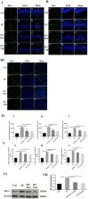
          1. The results of TUNEL staining showed that the expression in rats in the epilepsy group was the most obvious and was significantly different from that in rats in the control, EP+BAY and EP+TAK groups. 2. The expression of TLR4 and NF-κB was highest in rats in the epilepsy group and was significantly different from that in rats in the control, EP+BAY and EP+TAK groups. 3. The fluorescence intensity and number of IBA-1-positive cells in rats in the epilepsy group were highest and significantly different from those in rats in the control, EP+BAY and EP+TAK groups. Western blot analysis of IBA-1 showed that the expression in rats in the epilepsy group was the highest and was statistically significant. 4. CD68 was the highest in rats in the epilepsy group and was statistically significant. 5. In the open-field experiment, the central region residence time of rats in the EP group was delayed, the central region movement distance traveled was prolonged, the total distance traveled was prolonged, and the average speed was increased. Compared with rats in the EP group, rats in the EP+BAY and EP+ TAK groups exhibited improvements to different degrees.

          Conclusion

          At the tissue level, downregulation of the TLR4/NF-κB inflammatory pathway in epilepsy could inhibit microglial activation and the expression of the inflammatory factor CD68, could inhibit hyperphagocytosis, and inhibit the occurrence and exacerbation of epilepsy, thus improving cognitive and emotional disorders after epileptic seizures.

          Related collections

          Most cited references54

          • Record: found
          • Abstract: found
          • Article: not found

          ILAE classification of the epilepsies: Position paper of the ILAE Commission for Classification and Terminology

          The International League Against Epilepsy (ILAE) Classification of the Epilepsies has been updated to reflect our gain in understanding of the epilepsies and their underlying mechanisms following the major scientific advances that have taken place since the last ratified classification in 1989. As a critical tool for the practicing clinician, epilepsy classification must be relevant and dynamic to changes in thinking, yet robust and translatable to all areas of the globe. Its primary purpose is for diagnosis of patients, but it is also critical for epilepsy research, development of antiepileptic therapies, and communication around the world. The new classification originates from a draft document submitted for public comments in 2013, which was revised to incorporate extensive feedback from the international epilepsy community over several rounds of consultation. It presents three levels, starting with seizure type, where it assumes that the patient is having epileptic seizures as defined by the new 2017 ILAE Seizure Classification. After diagnosis of the seizure type, the next step is diagnosis of epilepsy type, including focal epilepsy, generalized epilepsy, combined generalized, and focal epilepsy, and also an unknown epilepsy group. The third level is that of epilepsy syndrome, where a specific syndromic diagnosis can be made. The new classification incorporates etiology along each stage, emphasizing the need to consider etiology at each step of diagnosis, as it often carries significant treatment implications. Etiology is broken into six subgroups, selected because of their potential therapeutic consequences. New terminology is introduced such as developmental and epileptic encephalopathy. The term benign is replaced by the terms self-limited and pharmacoresponsive, to be used where appropriate. It is hoped that this new framework will assist in improving epilepsy care and research in the 21st century.
            Bookmark
            • Record: found
            • Abstract: not found
            • Article: not found

            Modification of seizure activity by electrical stimulation: II. Motor seizure

              Bookmark
              • Record: found
              • Abstract: found
              • Article: found
              Is Open Access

              Brain-Derived Neurotrophic Factor in Brain Disorders: Focus on Neuroinflammation

              Brain-derived neurotrophic factor (BDNF) is one of the most studied neurotrophins in the healthy and diseased brain. As a result, there is a large body of evidence that associates BDNF with neuronal maintenance, neuronal survival, plasticity, and neurotransmitter regulation. Patients with psychiatric and neurodegenerative disorders often have reduced BDNF concentrations in their blood and brain. A current hypothesis suggests that these abnormal BDNF levels might be due to the chronic inflammatory state of the brain in certain disorders, as neuroinflammation is known to affect several BDNF-related signaling pathways. Activation of glia cells can induce an increase in the levels of pro- and antiinflammatory cytokines and reactive oxygen species, which can lead to the modulation of neuronal function and neurotoxicity observed in several brain pathologies. Understanding how neuroinflammation is involved in disorders of the brain, especially in the disease onset and progression, can be crucial for the development of new strategies of treatment. Despite the increasing evidence for the involvement of BDNF and neuroinflammation in brain disorders, there is scarce evidence that addresses the interaction between the neurotrophin and neuroinflammation in psychiatric and neurodegenerative diseases. This review focuses on the effect of acute and chronic inflammation on BDNF levels in the most common psychiatric and neurodegenerative disorders and aims to shed some light on the possible biological mechanisms that may influence this effect. In addition, this review will address the effect of behavior and pharmacological interventions on BDNF levels in these disorders.
                Bookmark

                Author and article information

                Contributors
                Journal
                Front Neurol
                Front Neurol
                Front. Neurol.
                Frontiers in Neurology
                Frontiers Media S.A.
                1664-2295
                14 April 2022
                2022
                : 13
                : 823908
                Affiliations
                Department of Pediatrics, Shengjing Hospital of China Medical University , Shenyang, China
                Author notes

                Edited by: Marino M. Bianchin, Federal University of Rio Grande Do Sul, Brazil

                Reviewed by: Yam Nath Paudel, Monash University Malaysia, Malaysia; Enes Akyuz, University of Health Sciences, Turkey

                *Correspondence: Hua Wang wanghcmu1663@ 123456163.com

                This article was submitted to Epilepsy, a section of the journal Frontiers in Neurology

                Article
                10.3389/fneur.2022.823908
                9046666
                35493845
                99010056-b210-4f67-a3ad-033fc50bae4d
                Copyright © 2022 Wu, Wang, Liu, Zhao and Zhang.

                This is an open-access article distributed under the terms of the Creative Commons Attribution License (CC BY). The use, distribution or reproduction in other forums is permitted, provided the original author(s) and the copyright owner(s) are credited and that the original publication in this journal is cited, in accordance with accepted academic practice. No use, distribution or reproduction is permitted which does not comply with these terms.

                History
                : 24 December 2021
                : 16 March 2022
                Page count
                Figures: 6, Tables: 0, Equations: 0, References: 54, Pages: 14, Words: 8222
                Categories
                Neurology
                Original Research

                Neurology
                epilepsy,microglial cells,brain injury,phagocytosis,emotional disorder,neurons
                Neurology
                epilepsy, microglial cells, brain injury, phagocytosis, emotional disorder, neurons

                Comments

                Comment on this article