16
views
0
recommends
+1 Recommend
0 collections
    0
    shares
      • Record: found
      • Abstract: found
      • Article: found
      Is Open Access

      Anaphase Chromosomes in Crane-Fly Spermatocytes Treated With Taxol (Paclitaxel) Accelerate When Their Kinetochore Microtubules Are Cut: Evidence for Spindle Matrix Involvement With Spindle Forces

      research-article

      Read this article at

      Bookmark
          There is no author summary for this article yet. Authors can add summaries to their articles on ScienceOpen to make them more accessible to a non-specialist audience.

          Abstract

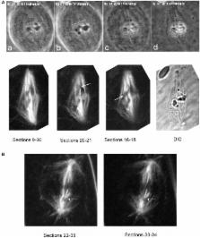
          Various experiments have indicated that anaphase chromosomes continue to move after their kinetochore microtubules are severed. The chromosomes move poleward at an accelerated rate after the microtubules are cut but they slow down 1–3 min later and move poleward at near the original speed. There are two published interpretations of chromosome movements with severed kinetochore microtubules. One interpretation is that dynein relocates to the severed microtubule ends and propels them poleward by pushing against non-kinetochore microtubules. The other interpretation is that components of a putative “spindle matrix” normally push kinetochore microtubules poleward and continue to do so after the microtubules are severed from the pole. In this study we distinguish between these interpretations by treating cells with taxol. Taxol eliminates microtubule dynamics, alters spindle microtubule arrangements, and inhibits dynein motor activity in vivo. If the dynein interpretation is correct, taxol should interfere with chromosome movements after kinetochore microtubules are severed because it alters the arrangements of spindle microtubules and because it blocks dynein activity. If the “spindle matrix” interpretation is correct, on the other hand, taxol should not interfere with the accelerated movements. Our results support the spindle matrix interpretation: anaphase chromosomes in taxol-treated crane-fly spermatocytes accelerated after their kinetochore microtubules were severed.

          Related collections

          Most cited references46

          • Record: found
          • Abstract: found
          • Article: not found

          Taxol suppresses dynamics of individual microtubules in living human tumor cells.

          Microtubules are intrinsically dynamic polymers, and their dynamics play a crucial role in mitotic spindle assembly, the mitotic checkpoint, and chromosome movement. We hypothesized that, in living cells, suppression of microtubule dynamics is responsible for the ability of taxol to inhibit mitotic progression and cell proliferation. Using quantitative fluorescence video microscopy, we examined the effects of taxol (30-100 nM) on the dynamics of individual microtubules in two living human tumor cell lines: Caov-3 ovarian adenocarcinoma cells and A-498 kidney carcinoma cells. Taxol accumulated more in Caov-3 cells than in A-498 cells. At equivalent intracellular taxol concentrations, dynamic instability was inhibited similarly in the two cell lines. Microtubule shortening rates were inhibited in Caov-3 cells and in A-498 cells by 32 and 26%, growing rates were inhibited by 24 and 18%, and dynamicity was inhibited by 31 and 63%, respectively. All mitotic spindles were abnormal, and many interphase cells became multinucleate (Caov-3, 30%; A-498, 58%). Taxol blocked cell cycle progress at the metaphase/anaphase transition and inhibited cell proliferation. The results indicate that suppression of microtubule dynamics by taxol deleteriously affects the ability of cancer cells to properly assemble a mitotic spindle, pass the metaphase/anaphase checkpoint, and produce progeny.
            Bookmark
            • Record: found
            • Abstract: found
            • Article: not found

            Insights into the mechanism of microtubule stabilization by Taxol.

            The antitumor drug Taxol stabilizes microtubules and reduces their dynamicity, promoting mitotic arrest and cell death. Upon assembly of the alpha/beta-tubulin heterodimer, GTP bound to beta-tubulin is hydrolyzed to GDP reaching a steady-state equilibrium between free tubulin dimers and microtubules. The binding of Taxol to beta-tubulin in the polymer results in cold-stable microtubules at the expense of tubulin dimers, even in the absence of exogenous GTP. However, there is little biochemical insight into the mechanism(s) by which Taxol stabilizes microtubules. Here, we analyze the structural changes occurring in both beta- and alpha-tubulin upon microtubule stabilization by Taxol. Hydrogen/deuterium exchange (HDX) coupled to liquid chromatography-electrospray ionization MS demonstrated a marked reduction in deuterium incorporation in both beta-and alpha-tubulin when Taxol was present. Decreased local HDX in peptic peptides was mapped on the tubulin structure and revealed both expected and new dimer-dimer interactions. The increased rigidity in Taxol microtubules was distinct from and complementary to that due to GTP-induced polymerization. The Taxol-induced changes in tubulin conformation act against microtubule depolymerization in a precise directional way. These results demonstrate that HDX coupled to liquid chromatography-electrospray ionization MS can be effectively used to study conformational effects induced by small ligands on microtubules. The present study also opens avenues for locating drug and protein binding sites and for deciphering the mechanisms by which their interactions alter the conformation of microtubules and tubulin dimers.
              Bookmark
              • Record: found
              • Abstract: found
              • Article: not found

              Repeat motifs of tau bind to the insides of microtubules in the absence of taxol.

              The tau family of microtubule-associated proteins has a microtubule-binding domain which includes three or four conserved sequence repeats. Pelleting assays show that when tubulin and tau are co- assembled into microtubules, the presence of taxol reduces the amount of tau incorporated. In the absence of taxol, strong binding sites for tau are filled by one repeat motif per tubulin dimer; additional tau molecules bind more weakly. We have labelled a repeat motif with nanogold and used three-dimensional electron cryomicroscopy to compare images of microtubules assembled with labelled or unlabelled tau. With kinesin motor domains bound to the microtubule outer surface to distinguish between alpha- and beta-tubulin, we show that the gold label lies on the inner surface close to the taxol binding site on beta-tubulin. Loops within the repeat motifs of tau have sequence similarity to an extended loop which occupies a site in alpha-tubulin equivalent to the taxol-binding pocket in beta-tubulin. We propose that loops in bound tau stabilize microtubules in a similar way to taxol, although with lower affinity so that assembly is reversible.
                Bookmark

                Author and article information

                Contributors
                Journal
                Front Cell Dev Biol
                Front Cell Dev Biol
                Front. Cell Dev. Biol.
                Frontiers in Cell and Developmental Biology
                Frontiers Media S.A.
                2296-634X
                24 July 2018
                2018
                : 6
                : 77
                Affiliations
                [1] 1Biology Department, York University , North York, ON, Canada
                [2] 2Beckman Laser Institute and Department of Biomedical Engineering, University of California, Irvine , Irvine, CA, United States
                [3] 3Department of Bioengineering and Institute for Engineering in Medicine, University of California, San Diego , San Diego, CA, United States
                Author notes

                Edited by: Daniel Leslie Fisher, Centre National de la Recherche Scientifique (CNRS), France

                Reviewed by: Song-Tao Liu, University of Toledo, United States; Andrew Burgess, Anzac Research Institute, Australia

                *Correspondence: Arthur Forer aforer@ 123456yorku.ca

                †Present Address: Rozhan Sheykhani, Department of Biology, Lakeland Community College, Kirtland, OH, United States

                This article was submitted to Cell Growth and Division, a section of the journal Frontiers in Cell and Developmental Biology

                Article
                10.3389/fcell.2018.00077
                6066604
                30087895
                8fe1bf9a-70c2-4cb1-b106-1cb5d9b8c6b6
                Copyright © 2018 Forer, Sheykhani and Berns.

                This is an open-access article distributed under the terms of the Creative Commons Attribution License (CC BY). The use, distribution or reproduction in other forums is permitted, provided the original author(s) and the copyright owner(s) are credited and that the original publication in this journal is cited, in accordance with accepted academic practice. No use, distribution or reproduction is permitted which does not comply with these terms.

                History
                : 08 May 2018
                : 29 June 2018
                Page count
                Figures: 8, Tables: 1, Equations: 0, References: 52, Pages: 11, Words: 6957
                Funding
                Funded by: Natural Sciences and Engineering Research Council of Canada 10.13039/501100000038
                Funded by: U.S. Air Force 10.13039/100006831
                Award ID: FA9550-17-1-0193
                Categories
                Cell and Developmental Biology
                Original Research

                mitotic mechanisms,spindle matrix,taxol,paclitaxel,laser irradiation,crane-fly spermatocytes

                Comments

                Comment on this article