24
views
0
recommends
+1 Recommend
0 collections
    0
    shares
      • Record: found
      • Abstract: found
      • Article: not found

      Resolution of infection promotes a state of dormancy and long survival of CD4 memory T cells

      research-article

      Read this article at

      ScienceOpenPublisherPMC
      Bookmark
          There is no author summary for this article yet. Authors can add summaries to their articles on ScienceOpen to make them more accessible to a non-specialist audience.

          Abstract

          Memory T cells survive throughout the lifetime of an individual and are protective upon recall. It is not clear how memory T cells can live so long. Here, we demonstrate that at the resolution of a viral infection, low levels of antigen are captured by B cells and presented to specific CD4 + memory T cells to render a state of unresponsiveness. We demonstrate in two systems that this process occurs naturally during the fall of antigen and is associated with a global gene expression program initiated with the clearance of antigen. Our study suggests that in the absence of antigen, a state of dormancy associated with low energy utilization and proliferation can help memory CD4 + T cells to survive nearly throughout the lifetime of mice. The dormant CD4 + memory T cells become activated by stimulatory signals generated by a subsequent infection. We propose that quiescence might be a mechanism necessary to regulate long-term survival of CD4 memory T cells and to prevent cross-reactivity to self, hence autoimmunity.

          Related collections

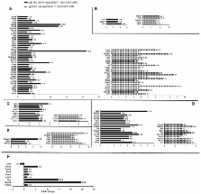
          Most cited references54

          • Record: found
          • Abstract: found
          • Article: not found

          CD4+ T cells are required for secondary expansion and memory in CD8+ T lymphocytes.

          A long-standing paradox in cellular immunology concerns the conditional requirement for CD4+ T-helper (T(H)) cells in the priming of cytotoxic CD8+ T lymphocyte (CTL) responses in vivo. Whereas CTL responses against certain viruses can be primed in the absence of CD4+ T cells, others, such as those mediated through 'cross-priming' by host antigen-presenting cells, are dependent on T(H) cells. A clearer understanding of the contribution of T(H) cells to CTL development has been hampered by the fact that most T(H)-independent responses have been demonstrated ex vivo as primary cytotoxic effectors, whereas T(H)-dependent responses generally require secondary in vitro re-stimulation for their detection. Here, we have monitored the primary and secondary responses of T(H)-dependent and T(H)-independent CTLs and find in both cases that CD4+ T cells are dispensable for primary expansion of CD8+ T cells and their differentiation into cytotoxic effectors. However, secondary CTL expansion (that is, a secondary response upon re-encounter with antigen) is wholly dependent on the presence of T(H) cells during, but not after, priming. Our results demonstrate that T-cell help is 'programmed' into CD8+ T cells during priming, conferring on these cells a hallmark of immune response memory: the capacity for functional expansion on re-encounter with antigen.
            Bookmark
            • Record: found
            • Abstract: found
            • Article: not found

            Effector and memory CTL differentiation.

            Technological advances in recent years have allowed for an ever-expanding ability to analyze and quantify in vivo immune responses. MHC tetramers, intracellular cytokine staining, an increasing repertoire of transgenic and "knockout" mice, and the detailed characterization of a variety of infectious models have all facilitated more precise and definitive analyses of the generation and function of cytotoxic T lymphocytes (CTL). Understanding the mechanisms behind the differentiation of effector and memory CTL is of increasing importance to develop vaccination strategies against a variety of established and emerging infectious diseases. This review focuses on recent advances in our understanding of how effector and memory CTL differentiate and survive in vivo in response to viral or bacterial infection.
              Bookmark
              • Record: found
              • Abstract: found
              • Article: not found

              Duration of antiviral immunity after smallpox vaccination.

              Although naturally occurring smallpox was eliminated through the efforts of the World Health Organization Global Eradication Program, it remains possible that smallpox could be intentionally released. Here we examine the magnitude and duration of antiviral immunity induced by one or more smallpox vaccinations. We found that more than 90% of volunteers vaccinated 25-75 years ago still maintain substantial humoral or cellular immunity (or both) against vaccinia, the virus used to vaccinate against smallpox. Antiviral antibody responses remained stable between 1-75 years after vaccination, whereas antiviral T-cell responses declined slowly, with a half-life of 8-15 years. If these levels of immunity are considered to be at least partially protective, then the morbidity and mortality associated with an intentional smallpox outbreak would be substantially reduced because of pre-existing immunity in a large number of previously vaccinated individuals.
                Bookmark

                Author and article information

                Journal
                8706300
                4179
                Immunol Cell Biol
                Immunology and cell biology
                0818-9641
                1440-1711
                27 December 2010
                1 March 2011
                November 2011
                1 May 2012
                : 89
                : 8
                : 870-881
                Affiliations
                [1 ]Department of Pathology, Johns Hopkins University, Baltimore MD 21205 USA
                [2 ]Department of Oncology, Johns Hopkins University, Baltimore MD 21205 USA
                [3 ]JHMI Microarray Core, Johns Hopkins University, Baltimore MD 21205 USA
                Author notes
                [$ ]Author for correspondence: Tel: 410-614-4931, Fax: 410-614-3548, ssadegh@ 123456jhmi.edu
                [*]

                equal contribution

                [#]

                Present address: Walter Reed Army Institute of Research, 503 Robert Grant Ave 3W110, Silver Spring MD 20910

                Article
                nihpa260641
                10.1038/icb.2011.2
                3131418
                21358746
                8306a972-4976-4aec-b401-3deaad651b7a

                Users may view, print, copy, download and text and data- mine the content in such documents, for the purposes of academic research, subject always to the full Conditions of use: http://www.nature.com/authors/editorial_policies/license.html#terms

                History
                Funding
                Funded by: National Institute of Allergy and Infectious Diseases Extramural Activities : NIAID
                Funded by: National Institute of General Medical Sciences : NIGMS
                Award ID: R56 AI063764-01A1 ||AI
                Funded by: National Institute of Allergy and Infectious Diseases Extramural Activities : NIAID
                Funded by: National Institute of General Medical Sciences : NIGMS
                Award ID: R01 GM053549-09 ||GM
                Funded by: National Institute of Allergy and Infectious Diseases Extramural Activities : NIAID
                Funded by: National Institute of General Medical Sciences : NIGMS
                Award ID: R01 GM053549-08 ||GM
                Funded by: National Institute of Allergy and Infectious Diseases Extramural Activities : NIAID
                Funded by: National Institute of General Medical Sciences : NIGMS
                Award ID: R01 AI063764-05S1 ||AI
                Funded by: National Institute of Allergy and Infectious Diseases Extramural Activities : NIAID
                Funded by: National Institute of General Medical Sciences : NIGMS
                Award ID: R01 AI063764-05 ||AI
                Funded by: National Institute of Allergy and Infectious Diseases Extramural Activities : NIAID
                Funded by: National Institute of General Medical Sciences : NIGMS
                Award ID: R01 AI063764-04 ||AI
                Funded by: National Institute of Allergy and Infectious Diseases Extramural Activities : NIAID
                Funded by: National Institute of General Medical Sciences : NIGMS
                Award ID: R01 AI063764-03 ||AI
                Funded by: National Institute of Allergy and Infectious Diseases Extramural Activities : NIAID
                Funded by: National Institute of General Medical Sciences : NIGMS
                Award ID: R01 AI063764-02 ||AI
                Funded by: National Institute of Allergy and Infectious Diseases Extramural Activities : NIAID
                Funded by: National Institute of General Medical Sciences : NIGMS
                Award ID: R01 AI063764-01A2 ||AI
                Categories
                Article

                Cell biology
                memory survival,memory t cells,b cell antigen presentation,memory t cells survival,gene regulation,low-dose antigen,microarray,anergy,bcr-mediated antigen capture,cd4 memory t cells

                Comments

                Comment on this article