58
views
0
recommends
+1 Recommend
0 collections
    0
    shares
      • Record: found
      • Abstract: found
      • Article: found
      Is Open Access

      Division of labour between Myc and G1 cyclins in cell cycle commitment and pace control

      research-article

      Read this article at

      Bookmark
          There is no author summary for this article yet. Authors can add summaries to their articles on ScienceOpen to make them more accessible to a non-specialist audience.

          Abstract

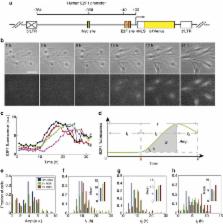
          A body of evidence has shown that the control of E2F transcription factor activity is critical for determining cell cycle entry and cell proliferation. However, an understanding of the precise determinants of this control, including the role of other cell-cycle regulatory activities, has not been clearly defined. Here, recognizing that the contributions of individual regulatory components could be masked by heterogeneity in populations of cells, we model the potential roles of individual components together with the use of an integrated system to follow E2F dynamics at the single-cell level and in real time. These analyses reveal that crossing a threshold amplitude of E2F accumulation determines cell cycle commitment. Importantly, we find that Myc is critical in modulating the amplitude, whereas cyclin D/E activities have little effect on amplitude but do contribute to the modulation of duration of E2F activation, thereby affecting the pace of cell cycle progression.

          Abstract

          The transcription factor E2F is critical for determining cell proliferation. By monitoring E2F activity in single cells throughout the cell cycle, Dong et al. provide evidence that Myc and G1 cyclin/CDKs regulate different aspects of E2F temporal dynamics, resulting in distinct phenotypic outputs.

          Related collections

          Most cited references40

          • Record: found
          • Abstract: found
          • Article: not found

          Encoding and decoding cellular information through signaling dynamics.

          A growing number of studies are revealing that cells can send and receive information by controlling the temporal behavior (dynamics) of their signaling molecules. In this Review, we discuss what is known about the dynamics of various signaling networks and their role in controlling cellular responses. We identify general principles that are emerging in the field, focusing specifically on how the identity and quantity of a stimulus is encoded in temporal patterns, how signaling dynamics influence cellular outcomes, and how specific dynamical patterns are both shaped and interpreted by the structure of molecular networks. We conclude by discussing potential functional roles for transmitting cellular information through the dynamics of signaling molecules and possible applications for the treatment of disease. Copyright © 2013 Elsevier Inc. All rights reserved.
            Bookmark
            • Record: found
            • Abstract: found
            • Article: not found

            p53 dynamics control cell fate.

            Cells transmit information through molecular signals that often show complex dynamical patterns. The dynamic behavior of the tumor suppressor p53 varies depending on the stimulus; in response to double-strand DNA breaks, it shows a series of repeated pulses. Using a computational model, we identified a sequence of precisely timed drug additions that alter p53 pulses to instead produce a sustained p53 response. This leads to the expression of a different set of downstream genes and also alters cell fate: Cells that experience p53 pulses recover from DNA damage, whereas cells exposed to sustained p53 signaling frequently undergo senescence. Our results show that protein dynamics can be an important part of a signal, directly influencing cellular fate decisions.
              Bookmark
              • Record: found
              • Abstract: found
              • Article: not found

              The proliferation-quiescence decision is controlled by a bifurcation in CDK2 activity at mitotic exit.

              Tissue homeostasis in metazoans is regulated by transitions of cells between quiescence and proliferation. The hallmark of proliferating populations is progression through the cell cycle, which is driven by cyclin-dependent kinase (CDK) activity. Here, we introduce a live-cell sensor for CDK2 activity and unexpectedly found that proliferating cells bifurcate into two populations as they exit mitosis. Many cells immediately commit to the next cell cycle by building up CDK2 activity from an intermediate level, while other cells lack CDK2 activity and enter a transient state of quiescence. This bifurcation is directly controlled by the CDK inhibitor p21 and is regulated by mitogens during a restriction window at the end of the previous cell cycle. Thus, cells decide at the end of mitosis to either start the next cell cycle by immediately building up CDK2 activity or to enter a transient G0-like state by suppressing CDK2 activity. Copyright © 2013 Elsevier Inc. All rights reserved.
                Bookmark

                Author and article information

                Journal
                Nat Commun
                Nat Commun
                Nature Communications
                Nature Pub. Group
                2041-1723
                01 September 2014
                : 5
                : 4750
                Affiliations
                [1 ]Computational Biology and Bioinformatics Program, Duke University , Durham, North Carolina 27708, USA
                [2 ]Department of Biomedical Engineering, Duke University , Durham, North Carolina 27708, USA
                [3 ]Department of Pharmacology and Cancer Biology, Duke University , Durham, North Carolina 27708, USA
                [4 ]Department of Molecular Genetics and Microbiology, Duke University , Durham, North Carolina 27708, USA
                [5 ]Department of Pediatrics, Duke University , Durham, North Carolina 27708, USA
                [6 ]Center for Genomic and Computational Biology, Duke University , Durham, North Carolina 27708, USA
                [7 ]Duke Center for Systems Biology, Duke University , Durham, North Carolina 27708, USA
                [8 ]Present address: School of Medicine, Johns Hopkins University, Baltimore, Maryland 21205, USA
                Author notes
                Article
                ncomms5750
                10.1038/ncomms5750
                4164785
                25175461
                7de9bb40-cabe-41db-8d9c-768bdf3dea72
                Copyright © 2014, Nature Publishing Group, a division of Macmillan Publishers Limited. All Rights Reserved.

                This work is licensed under a Creative Commons Attribution-NonCommercial-ShareAlike 4.0 International License. The images or other third party material in this article are included in the article’s Creative Commons license, unless indicated otherwise in the credit line; if the material is not included under the Creative Commons license, users will need to obtain permission from the license holder to reproduce the material. To view a copy of this license, visit http://creativecommons.org/licenses/by-nc-sa/4.0/

                History
                : 21 February 2014
                : 17 July 2014
                Categories
                Article

                Uncategorized
                Uncategorized

                Comments

                Comment on this article