35
views
0
recommends
+1 Recommend
0 collections
    0
    shares
      • Record: found
      • Abstract: found
      • Article: not found

      A transsynaptic nanocolumn aligns neurotransmitter release to receptors

      research-article

      Read this article at

      ScienceOpenPublisherPMC
      Bookmark
          There is no author summary for this article yet. Authors can add summaries to their articles on ScienceOpen to make them more accessible to a non-specialist audience.

          Abstract

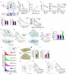
          Synaptic transmission is maintained by a delicate, subsynaptic molecular architecture, and even mild alterations in synapse structure drive functional changes during experience-dependent plasticity and pathological disorder 1, 2 . Key to this architecture is how the distribution of presynaptic vesicle fusion sites corresponds to the position of receptors in the postsynaptic density. However, despite long recognition that this spatial relationship modulates synaptic strength 3 , it has not been precisely described, due in part to the limited resolution of light microscopy. Using localization microscopy, we report here that key proteins mediating vesicle priming and fusion are mutually co-enriched within nanometer-scaled subregions of the presynaptic active zone. Through development of a new method to map vesicle fusion positions within single synapses, we found that action potential evoked fusion was guided by this protein gradient and occurred preferentially in confined areas with higher local density of RIM within the active zones. These presynaptic RIM nanoclusters closely aligned with concentrated postsynaptic receptors and scaffolding proteins 46 , suggesting a transsynaptic molecular “nanocolumn.” Thus, we propose that the nanoarchitecture of the active zone directs action potential evoked vesicle fusion to occur preferentially at sites directly opposing postsynaptic receptor-scaffold ensembles. Remarkably, NMDA receptor activation triggered distinct phases of plasticity in which postsynaptic reorganization was followed by transsynaptic nanoscale realignment. This architecture thus suggests a simple organizational principle of CNS synapses to maintain and modulate synaptic efficiency.

          Related collections

          Most cited references47

          • Record: found
          • Abstract: found
          • Article: not found

          Three-dimensional super-resolution imaging by stochastic optical reconstruction microscopy.

          Recent advances in far-field fluorescence microscopy have led to substantial improvements in image resolution, achieving a near-molecular resolution of 20 to 30 nanometers in the two lateral dimensions. Three-dimensional (3D) nanoscale-resolution imaging, however, remains a challenge. We demonstrated 3D stochastic optical reconstruction microscopy (STORM) by using optical astigmatism to determine both axial and lateral positions of individual fluorophores with nanometer accuracy. Iterative, stochastic activation of photoswitchable probes enables high-precision 3D localization of each probe, and thus the construction of a 3D image, without scanning the sample. Using this approach, we achieved an image resolution of 20 to 30 nanometers in the lateral dimensions and 50 to 60 nanometers in the axial dimension. This development allowed us to resolve the 3D morphology of nanoscopic cellular structures.
            Bookmark
            • Record: found
            • Abstract: found
            • Article: not found

            Direct stochastic optical reconstruction microscopy with standard fluorescent probes.

            Direct stochastic optical reconstruction microscopy (dSTORM) uses conventional fluorescent probes such as labeled antibodies or chemical tags for subdiffraction resolution fluorescence imaging with a lateral resolution of ∼20 nm. In contrast to photoactivated localization microscopy (PALM) with photoactivatable fluorescent proteins, dSTORM experiments start with bright fluorescent samples in which the fluorophores have to be transferred to a stable and reversible OFF state. The OFF state has a lifetime in the range of 100 milliseconds to several seconds after irradiation with light intensities low enough to ensure minimal photodestruction. Either spontaneously or photoinduced on irradiation with a second laser wavelength, a sparse subset of fluorophores is reactivated and their positions are precisely determined. Repetitive activation, localization and deactivation allow a temporal separation of spatially unresolved structures in a reconstructed image. Here we present a step-by-step protocol for dSTORM imaging in fixed and living cells on a wide-field fluorescence microscope, with standard fluorescent probes focusing especially on the photoinduced fine adjustment of the ratio of fluorophores residing in the ON and OFF states. Furthermore, we discuss labeling strategies, acquisition parameters, and temporal and spatial resolution. The ultimate step of data acquisition and data processing can be performed in seconds to minutes.
              Bookmark
              • Record: found
              • Abstract: found
              • Article: not found

              Superresolution imaging of chemical synapses in the brain.

              Determination of the molecular architecture of synapses requires nanoscopic image resolution and specific molecular recognition, a task that has so far defied many conventional imaging approaches. Here, we present a superresolution fluorescence imaging method to visualize the molecular architecture of synapses in the brain. Using multicolor, three-dimensional stochastic optical reconstruction microscopy, the distributions of synaptic proteins can be measured with nanometer precision. Furthermore, the wide-field, volumetric imaging method enables high-throughput, quantitative analysis of a large number of synapses from different brain regions. To demonstrate the capabilities of this approach, we have determined the organization of ten protein components of the presynaptic active zone and the postsynaptic density. Variations in synapse morphology, neurotransmitter receptor composition, and receptor distribution were observed both among synapses and across different brain regions. Combination with optogenetics further allowed molecular events associated with synaptic plasticity to be resolved at the single-synapse level. Copyright © 2010 Elsevier Inc. All rights reserved.
                Bookmark

                Author and article information

                Journal
                0410462
                6011
                Nature
                Nature
                Nature
                0028-0836
                1476-4687
                26 July 2016
                11 August 2016
                11 February 2017
                : 536
                : 7615
                : 210-214
                Affiliations
                [1 ]Department of Physiology, University of Maryland School of Medicine, Baltimore, MD 21201, USA
                [2 ]Program in Neuroscience, University of Maryland School of Medicine, Baltimore, MD 21201, USA
                [3 ]Medical Scientist Training Program, University of Maryland School of Medicine, Baltimore, MD 21201, USA
                [4 ]Department of Biology, Faculty of Science, Utrecht University, Utrecht, The Netherlands
                Author notes
                Correspondence and requests for materials should be addressed to T.A.B. ( tblanpied@ 123456som.umaryland.edu ) or A.T ( tangaihui@ 123456gmail.com )
                [*]

                These authors contributed equally to this work.

                Article
                NIHMS800441
                10.1038/nature19058
                5002394
                27462810
                7d82ac44-af1a-4c5e-bbc9-29e378b0968c

                Users may view, print, copy, and download text and data-mine the content in such documents, for the purposes of academic research, subject always to the full Conditions of use: http://www.nature.com/authors/editorial_policies/license.html#terms

                Reprints and permissions information is available at www.nature.com/reprints.

                History
                Categories
                Article

                Uncategorized
                Uncategorized

                Comments

                Comment on this article