6
views
0
recommends
+1 Recommend
0 collections
    0
    shares
      • Record: found
      • Abstract: found
      • Article: found
      Is Open Access

      Magnetic Resonance Imaging Versus Computed Tomography for Three‐Dimensional Bone Imaging of Musculoskeletal Pathologies: A Review

      review-article

      Read this article at

      Bookmark
          There is no author summary for this article yet. Authors can add summaries to their articles on ScienceOpen to make them more accessible to a non-specialist audience.

          Abstract

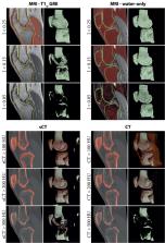
          Magnetic resonance imaging (MRI) is increasingly utilized as a radiation‐free alternative to computed tomography (CT) for the diagnosis and treatment planning of musculoskeletal pathologies. MR imaging of hard tissues such as cortical bone remains challenging due to their low proton density and short transverse relaxation times, rendering bone tissues as nonspecific low signal structures on MR images obtained from most sequences. Developments in MR image acquisition and post‐processing have opened the path for enhanced MR‐based bone visualization aiming to provide a CT‐like contrast and, as such, ease clinical interpretation. The purpose of this review is to provide an overview of studies comparing MR and CT imaging for diagnostic and treatment planning purposes in orthopedic care, with a special focus on selective bone visualization, bone segmentation, and three‐dimensional (3D) modeling. This review discusses conventional gradient‐echo derived techniques as well as dedicated short echo time acquisition techniques and post‐processing techniques, including the generation of synthetic CT, in the context of 3D and specific bone visualization. Based on the reviewed literature, it may be concluded that the recent developments in MRI‐based bone visualization are promising. MRI alone provides valuable information on both bone and soft tissues for a broad range of applications including diagnostics, 3D modeling, and treatment planning in multiple anatomical regions, including the skull, spine, shoulder, pelvis, and long bones.

          Level of Evidence

          3

          Technical Efficacy

          Stage 3

          Related collections

          Most cited references132

          • Record: found
          • Abstract: not found
          • Book Chapter: not found

          U-Net: Convolutional Networks for Biomedical Image Segmentation

            Bookmark
            • Record: found
            • Abstract: found
            • Article: not found

            Simple proton spectroscopic imaging.

            Simple modification of a spin echo imaging pulse sequence generates useful spectroscopic information at 0.35 T. New images are produced that show water only, fat only, and the difference between water and fat intensity. Imaging speed, spatial resolution, and signal-to-noise ratio are comparable with ordinary imaging. The method provides new parameters for tissue characterization and improved contrast between some organs.
              Bookmark
              • Record: found
              • Abstract: found
              • Article: not found

              Neuropsychological and Behavioral Outcomes after Exposure of Young Children to Procedures Requiring General Anesthesia

              Few studies of how exposure of children to anesthesia may affect neurodevelopment employ comprehensive neuropsychological assessments. This study tested the hypothesis that exposure to multiple, but not single, procedures requiring anesthesia before age 3 yr is associated with adverse neurodevelopmental outcomes.
                Bookmark

                Author and article information

                Contributors
                m.c.florkow@umcutrecht.nl
                p.seevinck@umcutrecht.nl
                Journal
                J Magn Reson Imaging
                J Magn Reson Imaging
                10.1002/(ISSN)1522-2586
                JMRI
                Journal of Magnetic Resonance Imaging
                John Wiley & Sons, Inc. (Hoboken, USA )
                1053-1807
                1522-2586
                19 January 2022
                July 2022
                : 56
                : 1 ( doiID: 10.1002/jmri.v56.1 )
                : 11-34
                Affiliations
                [ 1 ] Image Sciences Institute University Medical Center Utrecht Utrecht The Netherlands
                [ 2 ] Department of Orthopedics University Medical Center Utrecht Utrecht The Netherlands
                [ 3 ] Musculoskeletal Imaging Unit Imaging Center, Hospital da Luz Lisbon Portugal
                [ 4 ] Department of Radiology and Nuclear Medicine Erasmus MC, University Medical Center Rotterdam Rotterdam The Netherlands
                [ 5 ] MRIguidance BV Utrecht The Netherlands
                Author notes
                [*] [* ] Address reprint requests to: M.C.F. or P.R.S., Q.02.4.45, P.O. Box 85500, 3508 GA Utrecht, The Netherlands. E‐mail: m.c.florkow@ 123456umcutrecht.nl or p.seevinck@ 123456umcutrecht.nl

                Author information
                https://orcid.org/0000-0003-2520-7705
                https://orcid.org/0000-0002-8237-6321
                https://orcid.org/0000-0003-3727-3427
                Article
                JMRI28067
                10.1002/jmri.28067
                9305220
                35044717
                781f5204-be07-49d9-8a8d-b6900cf49c5f
                © 2022 The Authors. Journal of Magnetic Resonance Imaging published by Wiley Periodicals LLC on behalf of International Society for Magnetic Resonance in Medicine.

                This is an open access article under the terms of the http://creativecommons.org/licenses/by/4.0/ License, which permits use, distribution and reproduction in any medium, provided the original work is properly cited.

                History
                : 31 December 2021
                : 13 September 2021
                : 05 January 2022
                Page count
                Figures: 11, Tables: 2, Pages: 24, Words: 16100
                Funding
                Funded by: Stichting voor de Technische Wetenschappen , doi 10.13039/501100003958;
                Award ID: 15479
                Categories
                Review
                Reviews
                Custom metadata
                2.0
                July 2022
                Converter:WILEY_ML3GV2_TO_JATSPMC version:6.1.7 mode:remove_FC converted:21.07.2022

                Radiology & Imaging
                mri,ct,comparative review,diagnostic imaging,joints,bone and bones
                Radiology & Imaging
                mri, ct, comparative review, diagnostic imaging, joints, bone and bones

                Comments

                Comment on this article