31
views
0
recommends
+1 Recommend
0 collections
    0
    shares
      • Record: found
      • Abstract: found
      • Article: found
      Is Open Access

      Effect of Different Factors on Proliferation of Antler Cells, Cultured In Vitro

      research-article

      Read this article at

      Bookmark
          There is no author summary for this article yet. Authors can add summaries to their articles on ScienceOpen to make them more accessible to a non-specialist audience.

          Abstract

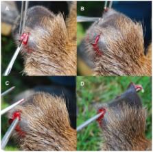
          Antlers as a potential model for bone growth and development have become an object of rising interest. To elucidate processes explaining how antler growth is regulated, in vitro cultures have been established. However, until now, there has been no standard method to cultivate antler cells and in vitro results are often opposite to those reported in vivo. In addition, many factors which are often not taken into account under in vitro conditions may play an important role in the development of antler cells. In this study we investigated the effects of the antler growth stage, the male individuality, passaged versus primary cultures and the effect of foetal calf serum concentrations on proliferative potential of mixed antler cell cultures in vitro, derived from regenerating antlers of red deer males ( Cervus elaphus). The proliferation potential of antler cells was measured by incorporation of 3H thymidine. Our results demonstrate that there is no significant effect of the antler growth stage, whereas male individuality and all other examined factors significantly affected antler cell proliferation. Furthermore, our results suggest that primary cultures may better represent in vivo conditions and processes occurring in regenerating antlers. In conclusion, before all main factors affecting antler cell proliferation in vitro will be satisfactorily investigated, results of in vitro studies focused on hormonal regulation of antler growth should be taken with extreme caution.

          Related collections

          Most cited references52

          • Record: found
          • Abstract: found
          • Article: not found

          Mesenchymal stem cells: progress toward promise.

          Despite having access to embryonic stem cells, many laboratories choose to study adult stem cells, not because of philosophical reasons but because of the practical aspects and day-to-day progress necessary for developing cellular therapeutics. There is certainly the ethical desire and responsibility to provide patients with therapies where few options exist. Multipotential cells have been isolated from adult tissues in many laboratories, characterized and their multipotentiality examined. Mesenchymal stem cells (MSC) can be isolated from several tissues but easily accessible BM seems to be the most common source. These adult stem cells may not be as 'powerful' or diverse as embryonic stem cells may one day become, but at present they offer many advantages for developing cellular therapeutics: ease of isolation, expansion potential, stable phenotype, shippability, and compatibility with different delivery methods and formulations. Their potential use as cellular therapeutics has prompted the investigation of interactions of allogeneic MSC with the immune response. The great importance of cardiovascular medicine has demanded that MSC also be tested in this discipline. We believe MSC continue to provide a substantial scientific and therapeutic opportunity, and have reviewed some of the recent developments in the field.
            Bookmark
            • Record: found
            • Abstract: found
            • Article: not found

            Serum deprivation of human marrow stromal cells (hMSCs) selects for a subpopulation of early progenitor cells with enhanced expression of OCT-4 and other embryonic genes.

            Recently there has been interest in developing cell and gene therapies with adult stem cells from human bone marrow referred to as mesenchymal stem cells or marrow stromal cells (hMSCs). We incubated early-passage hMSCs in serum-free medium without cytokines or other supplements for 2 to 4 weeks. Surprisingly, a subpopulation of the cells survived serum deprivation and then began to proliferate in serum-containing medium. The cells selected by serum deprivation had longer telomeres than control cells. Also, the patterns of gene expression revealed by reverse transcriptase-polymerase chain reaction (RT-PCR) assays and microarray data indicated that the cells selected by serum deprivation were a subpopulation of very early progenitor cells with enhanced expression of octomer-binding transcription factor 4 (OCT-4) and several other genes characteristically expressed in embryonic cells.
              Bookmark
              • Record: found
              • Abstract: found
              • Article: not found

              Deer antlers: a zoological curiosity or the key to understanding organ regeneration in mammals?

              Many organisms are able to regenerate lost or damaged body parts that are structural and functional replicates of the original. Eventually these become fully integrated into pre-existing tissues. However, with the exception of deer, mammals have lost this ability. Each spring deer shed antlers that were used for fighting and display during the previous mating season. Their loss is triggered by a fall in circulating testosterone levels, a hormonal change that is linked to an increase in day length. A complex 'blastema-like' structure or 'antler-bud' then forms; however, unlike the regenerative process in the newt, most evidence (albeit indirect) suggests that this does not involve reversal of the differentiated state but is stem cell based. The subsequent re-growth of antlers during the spring and summer months is spectacular and represents one of the fastest rates of organogenesis in the animal kingdom. Longitudinal growth involves endochondral ossification in the tip of each antler branch and bone growth around the antler shaft is by intramembranous ossification. As androgen concentrations rise in late summer, longitudinal growth stops, the skin (velvet) covering the antler is lost and antlers are 'polished' in preparation for the mating season. Although the timing of the antler growth cycle is clearly closely linked to circulating testosterone, oestrogen may be a key cellular regulator, as it is in the skeleton of other male mammals. We still know very little about the molecular machinery required for antler regeneration, although there is evidence that developmental signalling pathways with pleiotropic functions are important and that novel 'antler-specific' molecules may not exist. Identifying these pathways and factors, deciphering their interactions and how they are regulated by environmental cues could have an important impact on human health if this knowledge is applied to the engineering of new human tissues and organs.
                Bookmark

                Author and article information

                Contributors
                Role: Editor
                Journal
                PLoS One
                plos
                plosone
                PLoS ONE
                Public Library of Science (San Francisco, USA )
                1932-6203
                2011
                28 March 2011
                : 6
                : 3
                : e18053
                Affiliations
                [1 ]Department of Ethology, Institute of Animal Science, Prague - Uhříněves, Czech Republic
                [2 ]Department of Ecology, Charles University in Prague, Prague, Czech Republic
                [3 ]Department of Integrative Biology, University of Guelph, Guelph, Ontario, Canada
                The University of Hong Kong, Hong Kong
                Author notes

                Conceived and designed the experiments: EK LB. Performed the experiments: EK. Analyzed the data: EK LB. Contributed reagents/materials/analysis tools: RK. Wrote the paper: EK LB RK GAB.

                Article
                PONE-D-10-02521
                10.1371/journal.pone.0018053
                3065459
                21464927
                712794e5-210a-44be-aec0-36de0c0c9753
                Kužmová et al. This is an open-access article distributed under the terms of the Creative Commons Attribution License, which permits unrestricted use, distribution, and reproduction in any medium, provided the original author and source are credited.
                History
                : 16 September 2010
                : 24 February 2011
                Page count
                Pages: 6
                Categories
                Research Article
                Biology
                Anatomy and Physiology
                Musculoskeletal System
                Bone
                Cartilage
                Biotechnology
                Tissue Engineering
                Molecular Cell Biology
                Cellular Types
                Bone Marrow Cells
                Cell Growth
                Medicine
                Anatomy and Physiology
                Cell Physiology

                Uncategorized
                Uncategorized

                Comments

                Comment on this article