212
views
0
recommends
+1 Recommend
0 collections
    0
    shares
      • Record: found
      • Abstract: found
      • Article: found
      Is Open Access

      Autocrine Transforming Growth Factor-β1 Activation Mediated by Integrin αVβ3 Regulates Transcriptional Expression of Laminin-332 in Madin-Darby Canine Kidney Epithelial Cells

      research-article

      Read this article at

      Bookmark
          There is no author summary for this article yet. Authors can add summaries to their articles on ScienceOpen to make them more accessible to a non-specialist audience.

          Abstract

          The expression of the extracellular matrix protein Laminin-332 is regulated transcriptionally by TGF-β1 as a function of cell confluence in MDCK epithelial cells. Latent TGF-β1 is secreted apically, sequestered from its receptors and activation machinery, dependent on integrin αVβ3, localized on the basolateral side of the epithelial barrier.

          Abstract

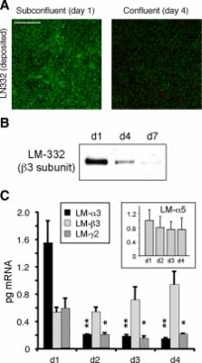
          Laminin (LM)-332 is an extracellular matrix protein that plays a structural role in normal tissues and is also important in facilitating recovery of epithelia from injury. We have shown that expression of LM-332 is up-regulated during renal epithelial regeneration after ischemic injury, but the molecular signals that control expression are unknown. Here, we demonstrate that in Madin-Darby canine kidney (MDCK) epithelial cells LM-332 expression occurs only in subconfluent cultures and is turned-off after a polarized epithelium has formed. Addition of active transforming growth factor (TGF)-β1 to confluent MDCK monolayers is sufficient to induce transcription of the LM α3 gene and LM-332 protein expression via the TGF-β type I receptor (TβR-I) and the Smad2–Smad4 complex. Significantly, we show that expression of LM-332 in MDCK cells is an autocrine response to endogenous TGF-β1 secretion and activation mediated by integrin αVβ3 because neutralizing antibodies block LM-332 production in subconfluent cells. In confluent cells, latent TGF-β1 is secreted apically, whereas TβR-I and integrin αVβ3 are localized basolaterally. Disruption of the epithelial barrier by mechanical injury activates TGF-β1, leading to LM-332 expression. Together, our data suggest a novel mechanism for triggering the production of LM-332 after epithelial injury.

          Related collections

          Most cited references74

          • Record: found
          • Abstract: found
          • Article: not found

          TGFbeta-SMAD signal transduction: molecular specificity and functional flexibility.

          Ligands of the transforming growth factor-beta (TGFbeta) superfamily of growth factors initiate signal transduction through a bewildering complexity of ligand-receptor interactions. Signalling then converges to nuclear accumulation of transcriptionally active SMAD complexes and gives rise to a plethora of specific functional responses in both embryos and adult organisms. Current research is focused on the mechanisms that regulate SMAD activity to evoke cell-type-specific and context-dependent transcriptional programmes. An equally important challenge is understanding the functional role of signal strength and duration. How are these quantitative aspects of the extracellular signal regulated? How are they then sensed and interpreted, and how do they affect responses?
            Bookmark
            • Record: found
            • Abstract: found
            • Article: not found

            From cells to organs: building polarized tissue.

            How do animal cells assemble into tissues and organs? A diverse array of tissue structures and shapes can be formed by organizing groups of cells into different polarized arrangements and by coordinating their polarity in space and time. Conserved design principles underlying this diversity are emerging from studies of model organisms and tissues. We discuss how conserved polarity complexes, signalling networks, transcription factors, membrane-trafficking pathways, mechanisms for forming lumens in tubes and other hollow structures, and transitions between different types of polarity, such as between epithelial and mesenchymal cells, are used in similar and iterative manners to build all tissues.
              Bookmark
              • Record: found
              • Abstract: found
              • Article: not found

              A simplified laminin nomenclature.

              A simplification of the laminin nomenclature is presented. Laminins are multidomain heterotrimers composed of alpha, beta and gamma chains. Previously, laminin trimers were numbered with Arabic numerals in the order discovered, that is laminins-1 to -5. We introduce a new identification system for a trimer using three Arabic numerals, based on the alpha, beta and gamma chain numbers. For example, the laminin with the chain composition alpha5beta1gamma1 is termed laminin-511, and not laminin-10. The current practice is also to mix two overlapping domain and module nomenclatures. Instead of the older Roman numeral nomenclature and mixed nomenclature, all modules are now called domains. Some domains are renamed or renumbered. Laminin epidermal growth factor-like (LE) domains are renumbered starting at the N-termini, to be consistent with general protein nomenclature. Domain IVb of alpha chains is named laminin 4a (L4a), domain IVa of alpha chains is named L4b, domain IV of gamma chains is named L4, and domain IV of beta chains is named laminin four (LF). The two coiled-coil domains I and II are now considered one laminin coiled-coil domain (LCC). The interruption in the coiled-coil of beta chains is named laminin beta-knob (Lbeta) domain. The chain origin of a domain is specified by the chain nomenclature, such as alpha1L4a. The abbreviation LM is suggested for laminin. Otherwise, the nomenclature remains unaltered.
                Bookmark

                Author and article information

                Contributors
                Role: Monitoring Editor
                Journal
                Mol Biol Cell
                mbc
                mbc
                Mol. Bio. Cell
                Molecular Biology of the Cell
                The American Society for Cell Biology
                1059-1524
                1939-4586
                1 November 2010
                : 21
                : 21
                : 3654-3668
                Affiliations
                [1]*Department of Surgery, Committee on Cell Physiology, and Committee on Molecular Pathogenesis and Molecular Medicine, The University of Chicago, Chicago, IL 60637; and
                [2] Institute for Oral and Musculoskeletal Biology, Dental School/Institute for Biochemistry II and Center for Molecular Medicine Cologne (CMMC) Medical Center, University of Cologne, D-50931 Cologne, Germany
                Author notes
                Address correspondence to: Jose V. Moyano ( jvmoyano@ 123456uchicago.edu ).
                Article
                3641173
                10.1091/mbc.E10-06-0523
                2965683
                20844080
                6bc9e954-8cf6-4db6-8c41-9a1c37d1d847
                © 2010 by The American Society for Cell Biology

                This article is distributed by The American Society for Cell Biology under license from the author(s). Two months after publication it is available to the public under an Attribution–Noncommercial–Share Alike 3.0 Unported Creative Commons License ( http://creativecommons.org/licenses/by-nc-sa/3.0).

                History
                : 21 June 2010
                : 23 August 2010
                : 2 September 2010
                Categories
                Articles
                Cell Interactions

                Molecular biology
                Molecular biology

                Comments

                Comment on this article