5
views
0
recommends
+1 Recommend
1 collections
    0
    shares
      • Record: found
      • Abstract: found
      • Article: found
      Is Open Access

      CT Examinations for COVID-19: A Systematic Review of Protocols, Radiation Dose, and Numbers Needed to Diagnose and Predict Translated title: COVID-19 진단을 위한 CT 검사: 프로토콜, 방사선량에 대한 체계적 문헌고찰 및 진단을 위한 CT 검사량

      research-article

      Read this article at

      Bookmark
          There is no author summary for this article yet. Authors can add summaries to their articles on ScienceOpen to make them more accessible to a non-specialist audience.

          Abstract

          Purpose

          Although chest CT has been discussed as a first-line test for coronavirus disease 2019 (COVID-19), little research has explored the implications of CT exposure in the population. To review chest CT protocols and radiation doses in COVID-19 publications and explore the number needed to diagnose (NND) and the number needed to predict (NNP) if CT is used as a first-line test.

          Materials and Methods

          We searched nine highly cited radiology journals to identify studies discussing the CT-based diagnosis of COVID-19 pneumonia. Study-level information on the CT protocol and radiation dose was collected, and the doses were compared with each national diagnostic reference level (DRL). The NND and NNP, which depends on the test positive rate (TPR), were calculated, given a CT sensitivity of 94% (95% confidence interval [CI]: 91%–96%) and specificity of 37% (95% CI: 26%–50%), and applied to the early outbreak in Wuhan, New York, and Italy.

          Results

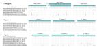
          From 86 studies, the CT protocol and radiation dose were reported in 81 (94.2%) and 17 studies (19.8%), respectively. Low-dose chest CT was used more than twice as often as standard-dose chest CT (39.5% vs.18.6%), while the remaining studies (44.2%) did not provide relevant information. The radiation doses were lower than the national DRLs in 15 of the 17 studies (88.2%) that reported doses. The NND was 3.2 scans (95% CI: 2.2–6.0). The NNPs at TPRs of 50%, 25%, 10%, and 5% were 2.2, 3.6, 8.0, 15.5 scans, respectively. In Wuhan, 35418 (TPR, 58%; 95% CI: 27710–56755) to 44840 (TPR, 38%; 95% CI: 35161–68164) individuals were estimated to have undergone CT examinations to diagnose 17365 patients. During the early surge in New York and Italy, daily NNDs changed up to 5.4 and 10.9 times, respectively, within 10 weeks.

          Conclusion

          Low-dose CT protocols were described in less than half of COVID-19 publications, and radiation doses were frequently lacking. The number of populations involved in a first-line diagnostic CT test could vary dynamically according to daily TPR; therefore, caution is required in future planning.

          Translated abstract

          목적

          Coronavirus disease 2019 (이하 COVID-19) 폐렴에서 CT를 일차 진단 검사로 사용하고자 하는 논의가 있지만, 대규모 인구에게 CT 검사를 적용했을 때의 상황을 고찰한 연구는 없었다. 본 연구에서는 COVID-19 폐렴을 다룬 연구들에서 CT 프로토콜과 방사선량을 분석하고, CT 검사가 일차 진단 검사법으로 사용될 때 필요한 CT 검사량에 대해 알아보고자 한다.

          대상과 방법

          본 연구는 9개의 인용도가 높은 영상의학과 저널에서 COVID-19 폐렴의 CT 기반 진단을 다룬 문헌들을 검색하였다. 먼저, 연구에서 제시된 CT 프로토콜, 방사선량을 조사하여, 이를 해당 국가의 diagnostic reference level과 비교하였다. 추가로, COVID-19에 대한 CT 민감도 94%, 특이도 37%를 적용하여, 우한시와 뉴욕, 이탈리아의 초기 COVID-19 outbreak에서 polymerase chain reaction (이하 PCR) 검사 양성률에 기반한 number needed to diagnose (이하 NND)와 number needed to predict (이하 NNP)를 계산하였다.

          결과

          총 86개의 연구가 검색되었고, 그중 CT 프로토콜은 81개의 연구에서(94.2%), 방사선량은 17개의 연구에서(19.8%) 보고되었다. 저선량 흉부 CT는 표준선량 흉부 CT보다 2배 많은 연구에서 활용되었다(39.5% vs. 18.6%). 방사선량을 보고한 17개의 연구들 중, 15개의 연구에서 방사선량은 해당 국가의 diagnostic reference level 수치보다 낮았다(88.2%). COVID-19에 대한 CT 민감도 94%, 특이도 37%를 적용하였을 때, NND는 3.2회 CT scans으로 나타났다. 한편, PCR 검사 양성률 50%, 25%, 10%, 5%에서의 한 명의 COVID-19 환자를 진단 위한 CT 검사량을 나타내는 NNP는 각각 2.2, 3.6, 8.0, 15.5회의 CT scans로 나타났다. 우한시에서는 최종 17365명의 COVID-19 환자를 진단하기 위하여 약 35418명에서(PCR 검사 양성률 58%) 44840명(PCR 검사 양성률 38%)의 사람들이 CT 검사를 받은 것으로 나타났다. 뉴욕시와 이탈리아의 초기 COVID-19 유행 10주간, PCR 검사 양성률에 따라 일 CT 검사량이 최대 5.4, 10.9배까지 변화하였다.

          결론

          CT를 COVID-19에 대한 일차적인 진단검사로 사용할 경우, PCR 검사 양성률에 따라 CT 검사량은 변동량이 크고, 이는 추후 판데믹 상황에서 고려되어야 할 것이다.

          Related collections

          Most cited references115

          • Record: found
          • Abstract: found
          • Article: not found

          Clinical Characteristics of Coronavirus Disease 2019 in China

          Abstract Background Since December 2019, when coronavirus disease 2019 (Covid-19) emerged in Wuhan city and rapidly spread throughout China, data have been needed on the clinical characteristics of the affected patients. Methods We extracted data regarding 1099 patients with laboratory-confirmed Covid-19 from 552 hospitals in 30 provinces, autonomous regions, and municipalities in mainland China through January 29, 2020. The primary composite end point was admission to an intensive care unit (ICU), the use of mechanical ventilation, or death. Results The median age of the patients was 47 years; 41.9% of the patients were female. The primary composite end point occurred in 67 patients (6.1%), including 5.0% who were admitted to the ICU, 2.3% who underwent invasive mechanical ventilation, and 1.4% who died. Only 1.9% of the patients had a history of direct contact with wildlife. Among nonresidents of Wuhan, 72.3% had contact with residents of Wuhan, including 31.3% who had visited the city. The most common symptoms were fever (43.8% on admission and 88.7% during hospitalization) and cough (67.8%). Diarrhea was uncommon (3.8%). The median incubation period was 4 days (interquartile range, 2 to 7). On admission, ground-glass opacity was the most common radiologic finding on chest computed tomography (CT) (56.4%). No radiographic or CT abnormality was found in 157 of 877 patients (17.9%) with nonsevere disease and in 5 of 173 patients (2.9%) with severe disease. Lymphocytopenia was present in 83.2% of the patients on admission. Conclusions During the first 2 months of the current outbreak, Covid-19 spread rapidly throughout China and caused varying degrees of illness. Patients often presented without fever, and many did not have abnormal radiologic findings. (Funded by the National Health Commission of China and others.)
            Bookmark
            • Record: found
            • Abstract: found
            • Article: found
            Is Open Access

            Detection of 2019 novel coronavirus (2019-nCoV) by real-time RT-PCR

            Background The ongoing outbreak of the recently emerged novel coronavirus (2019-nCoV) poses a challenge for public health laboratories as virus isolates are unavailable while there is growing evidence that the outbreak is more widespread than initially thought, and international spread through travellers does already occur. Aim We aimed to develop and deploy robust diagnostic methodology for use in public health laboratory settings without having virus material available. Methods Here we present a validated diagnostic workflow for 2019-nCoV, its design relying on close genetic relatedness of 2019-nCoV with SARS coronavirus, making use of synthetic nucleic acid technology. Results The workflow reliably detects 2019-nCoV, and further discriminates 2019-nCoV from SARS-CoV. Through coordination between academic and public laboratories, we confirmed assay exclusivity based on 297 original clinical specimens containing a full spectrum of human respiratory viruses. Control material is made available through European Virus Archive – Global (EVAg), a European Union infrastructure project. Conclusion The present study demonstrates the enormous response capacity achieved through coordination of academic and public laboratories in national and European research networks.
              Bookmark
              • Record: found
              • Abstract: found
              • Article: not found

              Correlation of Chest CT and RT-PCR Testing in Coronavirus Disease 2019 (COVID-19) in China: A Report of 1014 Cases

              Background Chest CT is used for diagnosis of 2019 novel coronavirus disease (COVID-19), as an important complement to the reverse-transcription polymerase chain reaction (RT-PCR) tests. Purpose To investigate the diagnostic value and consistency of chest CT as compared with comparison to RT-PCR assay in COVID-19. Methods From January 6 to February 6, 2020, 1014 patients in Wuhan, China who underwent both chest CT and RT-PCR tests were included. With RT-PCR as reference standard, the performance of chest CT in diagnosing COVID-19 was assessed. Besides, for patients with multiple RT-PCR assays, the dynamic conversion of RT-PCR results (negative to positive, positive to negative, respectively) was analyzed as compared with serial chest CT scans for those with time-interval of 4 days or more. Results Of 1014 patients, 59% (601/1014) had positive RT-PCR results, and 88% (888/1014) had positive chest CT scans. The sensitivity of chest CT in suggesting COVID-19 was 97% (95%CI, 95-98%, 580/601 patients) based on positive RT-PCR results. In patients with negative RT-PCR results, 75% (308/413) had positive chest CT findings; of 308, 48% were considered as highly likely cases, with 33% as probable cases. By analysis of serial RT-PCR assays and CT scans, the mean interval time between the initial negative to positive RT-PCR results was 5.1 ± 1.5 days; the initial positive to subsequent negative RT-PCR result was 6.9 ± 2.3 days). 60% to 93% of cases had initial positive CT consistent with COVID-19 prior (or parallel) to the initial positive RT-PCR results. 42% (24/57) cases showed improvement in follow-up chest CT scans before the RT-PCR results turning negative. Conclusion Chest CT has a high sensitivity for diagnosis of COVID-19. Chest CT may be considered as a primary tool for the current COVID-19 detection in epidemic areas. A translation of this abstract in Farsi is available in the supplement. - ترجمه چکیده این مقاله به فارسی، در ضمیمه موجود است.
                Bookmark

                Author and article information

                Journal
                Taehan Yongsang Uihakhoe Chi
                Taehan Yongsang Uihakhoe Chi
                JKSR
                Journal of the Korean Society of Radiology (Taehan Yŏngsang Ŭihakhoe chi)
                The Korean Society of Radiology
                1738-2637
                2288-2928
                November 2021
                04 November 2021
                : 82
                : 6
                : 1505-1523
                Affiliations
                [1 ]Department of Radiology, Seoul National University Hospital, Seoul National University College of Medicine, Seoul, Korea. [1 ]서울대학교 의과대학 서울대학교병원 영상의학과
                [2 ]Medical Research Collaborating Center, Seoul National University Hospital, Seoul, Korea. [2 ]서울대학교병원 의학연구협력센터
                [3 ]Department of Radiology, UMass Memorial Medical Center, Worcester, MA, USA. [3 ]Department of Radiology, UMass Memorial Medical Center
                Author notes
                Corresponding author: Soon Ho Yoon, MD. Department of Radiology, Seoul National University Hospital, Seoul National University College of Medicine, 101 Daehak-ro, Jongno-gu, Seoul 03080, Korea; Department of Radiology, UMass Memorial Medical Center, 55 Lake Avenue North, Worcester, MA 01655, USA. Tel 82-2-2072-2254, Fax 82-2-743-6385, yshoka@ 123456gmail.com
                Article
                10.3348/jksr.2021.0096
                9431975
                669c951f-3492-43d4-ac4b-9839aba3722e
                Copyrights © 2021 The Korean Society of Radiology

                This is an Open Access article distributed under the terms of the Creative Commons Attribution Non-Commercial License ( https://creativecommons.org/licenses/by-nc/4.0) which permits unrestricted non-commercial use, distribution, and reproduction in any medium, provided the original work is properly cited.

                History
                : 21 May 2021
                : 25 July 2021
                : 28 July 2021
                Categories
                Thoracic Imaging
                Original Article

                coronavirus,tomography, x-ray computed,radiation dosage,mass screening,clinical protocols

                Comments

                Comment on this article