71
views
0
recommends
+1 Recommend
0 collections
    0
    shares
      • Record: found
      • Abstract: found
      • Article: not found

      Exploration of Type II Binding Mode: A Privileged Approach for Kinase Inhibitor Focused Drug Discovery?

      review-article

      Read this article at

      ScienceOpenPublisherPMC
      Bookmark
          There is no author summary for this article yet. Authors can add summaries to their articles on ScienceOpen to make them more accessible to a non-specialist audience.

          Abstract

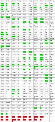
          The ATP site of kinases displays remarkable conformational flexibility when accommodating chemically diverse small molecule inhibitors. The so-called activation segment, whose conformation controls catalytic activity and access to the substrate binding pocket, can undergo a large conformational change with the active state assuming a ‘DFG-in’ and an inactive state assuming a ‘DFG-out’ conformation. Compounds that preferentially bind to the DFG-out conformation are typically called ‘type II’ inhibitors in contrast to ‘type I’ inhibitors that bind to the DFG-in conformation. This review surveys the large number of type II inhibitors that have been developed and provides an analysis of their crystallographically determined binding modes. Using a small library of type II inhibitors, we demonstrate that more than 200 kinases can be targeted, suggesting that type II inhibitors may not be intrinsically more selective than type I inhibitors.

          Related collections

          Most cited references46

          • Record: found
          • Abstract: found
          • Article: not found

          A small molecule-kinase interaction map for clinical kinase inhibitors.

          Kinase inhibitors show great promise as a new class of therapeutics. Here we describe an efficient way to determine kinase inhibitor specificity by measuring binding of small molecules to the ATP site of kinases. We have profiled 20 kinase inhibitors, including 16 that are approved drugs or in clinical development, against a panel of 119 protein kinases. We find that specificity varies widely and is not strongly correlated with chemical structure or the identity of the intended target. Many novel interactions were identified, including tight binding of the p38 inhibitor BIRB-796 to an imatinib-resistant variant of the ABL kinase, and binding of imatinib to the SRC-family kinase LCK. We also show that mutations in the epidermal growth factor receptor (EGFR) found in gefitinib-responsive patients do not affect the binding affinity of gefitinib or erlotinib. Our results represent a systematic small molecule-protein interaction map for clinical compounds across a large number of related proteins.
            Bookmark
            • Record: found
            • Abstract: found
            • Article: not found

            Structural mechanism for STI-571 inhibition of abelson tyrosine kinase.

            The inadvertent activation of the Abelson tyrosine kinase (Abl) causes chronic myelogenous leukemia (CML). A small-molecule inhibitor of Abl (STI-571) is effective in the treatment of CML. We report the crystal structure of the catalytic domain of Abl, complexed to a variant of STI-571. Critical to the binding of STI-571 is the adoption by the kinase of an inactive conformation, in which a centrally located "activation loop" is not phosphorylated. The conformation of this loop is distinct from that in active protein kinases, as well as in the inactive form of the closely related Src kinases. These results suggest that compounds that exploit the distinctive inactivation mechanisms of individual protein kinases can achieve both high affinity and high specificity.
              Bookmark
              • Record: found
              • Abstract: found
              • Article: not found

              Rational design of inhibitors that bind to inactive kinase conformations.

              The majority of kinase inhibitors that have been developed so far--known as type I inhibitors--target the ATP binding site of the kinase in its active conformation, in which the activation loop is phosphorylated. Recently, crystal structures of inhibitors such as imatinib (STI571), BIRB796 and sorafenib (BAY43-9006)--known as type II inhibitors--have revealed a new binding mode that exploits an additional binding site immediately adjacent to the region occupied by ATP. This pocket is made accessible by an activation-loop rearrangement that is characteristic of kinases in an inactive conformation. Here, we present a structural analysis of binding modes of known human type II inhibitors and demonstrate that they conform to a pharmacophore model that is currently being used to design a new generation of kinase inhibitors.
                Bookmark

                Author and article information

                Journal
                ACS Chem Biol
                ACS Chem. Biol
                cb
                acbcct
                ACS Chemical Biology
                American Chemical Society
                1554-8929
                1554-8937
                14 April 2014
                20 June 2014
                : 9
                : 6
                : 1230-1241
                Affiliations
                []High Magnetic Field Laboratory, Chinese Academy of Sciences, P.O. Box 1110, Hefei, Anhui 230031, P. R. China
                []University of Science and Technology of China , Hefei, Anhui 230036, P. R. China
                [§ ]Wellspring Biosciences LLC, 3210 Merryfield Row, San Diego, California 92121, United States
                []Structural Genomics Consortium, University of Oxford , Old Road Campus Research Building, Roosevelt Drive, Oxford OX3 7DQ, United Kingdom
                []Target Discovery Institute, University of Oxford , NDM Research Building, Roosevelt Drive, Oxford OX3 7LD, United Kingdom
                []Department of Biological Chemistry and Molecular Pharmacology, Harvard Medical School , 250 Longwood Avenue, Boston, Massachussetts 02115, United States
                Author notes
                Article
                10.1021/cb500129t
                4068218
                24730530
                5d470cc5-272a-4570-9d73-ce22ef8a8146
                Copyright © 2014 American Chemical Society

                Terms of Use CC-BY

                History
                : 19 February 2014
                : 14 April 2014
                Categories
                Reviews
                Custom metadata
                cb500129t
                cb-2014-00129t

                Biochemistry
                Biochemistry

                Comments

                Comment on this article