13
views
0
recommends
+1 Recommend
0 collections
    0
    shares
      • Record: found
      • Abstract: found
      • Article: found
      Is Open Access

      Gafchromic EBT3 film dosimetry in electron beams — energy dependence and improved film read‐out

      research-article

      Read this article at

      Bookmark
          There is no author summary for this article yet. Authors can add summaries to their articles on ScienceOpen to make them more accessible to a non-specialist audience.

          Abstract

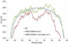
          For megavoltage photon radiation, the fundamental dosimetry characteristics of Gafchromic EBT3 film were determined in 60 Co gamma ray beam with addition of experimental and Monte Carlo (MC)‐simulated energy dependence of the film for 6 MV photon beam and 6 MeV, 9 MeV, 12 MeV, and 16 MeV electron beams in water phantom. For the film read‐out, two phase correction of scanner sensitivity was applied: a matrix correction for scanning area and dose‐dependent correction by iterative procedure. With these corrections, the uniformity of response can be improved to be within ± 50 pixel values (PVs). To improve the read‐out accuracy, a procedure with flipped film orientations was established. With the method, scanner uniformity can be improved further and dust particles, scratches and/or dirt on scanner glass can be detected and eliminated. Responses from red and green channels were averaged for read‐out, which decreased the effect of noise present in values from separate channels. Since the signal level with the blue channel is considerably lower than with other channels, the signal variation due to different perturbation effects increases the noise level so that the blue channel is not recommended to be used for dose determination. However, the blue channel can be used for the detection of emulsion thickness variations for film quality evaluations with unexposed films. With electron beams ranging from 6 MeV to 16 MeV and at reference measurement conditions in water, the energy dependence of the EBT3 film is uniform within 0.5%, with uncertainties close to 1.6% ( k = 2 ) . Including 6 MV photon beam and the electron beams mentioned, the energy dependence is within 1.1%. No notable differences were found between the experimental and MC‐simulated responses, indicating negligible change in intrinsic energy dependence of the EBT3 film for 6 MV photon beam and 6 MeV–16 MeV electron beams. Based on the dosimetric characteristics of the EBT3 film, the read‐out procedure established, the nearly uniform energy dependence found and the estimated uncertainties, the EBT3 film was concluded to be a suitable 2D dosimeter for measuring electron or mixed photon/electron dose distributions in water phantom. Uncertainties of 3.7% ( k = 2 ) for absolute and 2.3% ( k = 2 ) for relative dose were estimated.

          PACS numbers: 87.53.Bn, 87.55.K‐, 87.55.Qr

          Related collections

          Most cited references33

          • Record: found
          • Abstract: found
          • Article: not found

          BEAM: a Monte Carlo code to simulate radiotherapy treatment units.

          This paper describes BEAM, a general purpose Monte Carlo code to simulate the radiation beams from radiotherapy units including high-energy electron and photon beams, 60Co beams and orthovoltage units. The code handles a variety of elementary geometric entities which the user puts together as needed (jaws, applicators, stacked cones, mirrors, etc.), thus allowing simulation of a wide variety of accelerators. The code is not restricted to cylindrical symmetry. It incorporates a variety of powerful variance reduction techniques such as range rejection, bremsstrahlung splitting and forcing photon interactions. The code allows direct calculation of charge in the monitor ion chamber. It has the capability of keeping track of each particle's history and using this information to score separate dose components (e.g., to determine the dose from electrons scattering off the applicator). The paper presents a variety of calculated results to demonstrate the code's capabilities. The calculated dose distributions in a water phantom irradiated by electron beams from the NRC 35 MeV research accelerator, a Varian Clinac 2100C, a Philips SL75-20, an AECL Therac 20 and a Scanditronix MM50 are all shown to be in good agreement with measurements at the 2 to 3% level. Eighteen electron spectra from four different commercial accelerators are presented and various aspects of the electron beams from a Clinac 2100C are discussed. Timing requirements and selection of parameters for the Monte Carlo calculations are discussed.
            Bookmark
            • Record: found
            • Abstract: found
            • Article: not found

            Multichannel film dosimetry with nonuniformity correction.

            A new method to evaluate radiochromic film dosimetry data scanned in multiple color channels is presented. This work was undertaken to demonstrate that the multichannel method is fundamentally superior to the traditional single channel method. The multichannel method allows for the separation and removal of the nondose-dependent portions of a film image leaving a residual image that is dependent only on absorbed dose. Radiochromic films were exposed to 10 x 10 cm radiation fields (Co-60 and 6 MV) at doses up to about 300 cGy. The films were scanned in red-blue-green (RGB) format on a flatbed color scanner and measured to build calibration tables relating the absorbed dose to the response of the film in each of the color channels. Film images were converted to dose maps using two methods. The first method used the response from a single color channel and the second method used the response from all three color channels. The multichannel method allows for the separation of the scanned signal into one part that is dose-dependent and another part that is dose-independent and enables the correction of a variety of disturbances in the digitized image including nonuniformities in the active coating on the radiochromic film as well as scanner related artifacts. The fundamental mathematics of the two methods is described and the dose maps calculated from film images using the two methods are compared and analyzed. The multichannel dosimetry method was shown to be an effective way to separate out non-dose-dependent abnormalities from radiochromic dosimetry film images. The process was shown to remove disturbances in the scanned images caused by nonhomogeneity of the radiochromic film and artifacts caused by the scanner and to improve the integrity of the dose information. Multichannel dosimetry also reduces random noise in the dose images and mitigates scanner-related artifacts such as lateral position dependence. In providing an ability to calculate dose maps from data in all the color channels the multichannel method provides the ability to examine the agreement between the color channels. Furthermore, when using calibration data to convert RGB film images to dose using the new method, poor correspondence between the dose calculations for the three color channels provides an important indication that the this new technique enables easy indication in case the dose and calibration films are curve mismatched. The method permit compensation for thickness nonuniformities in the film, increases the signal to noise level, mitigates the lateral dose-dependency of flatbed scanners effect of the calculated dose map and extends the evaluable dose range to 10 cGy-100 Gy. Multichannel dosimetry with radiochromic film like Gafchromic EBT2 is shown to have significant advantages over single channel dosimetry. It is recommended that the dosimetry protocols described be implemented when using this radiochromic film to ensure the best data integrity and dosimetric accuracy.
              Bookmark
              • Record: found
              • Abstract: found
              • Article: not found

              An efficient protocol for radiochromic film dosimetry combining calibration and measurement in a single scan.

              Radiochromic film provides dose measurement at high spatial resolution, but often is not preferred for routine evaluation of patient-specific intensity modulated radiation therapy (IMRT) plans owing to ease-of-use factors. The authors have established an efficient protocol that combines calibration and measurement in a single scan and enables measurement results to be obtained in less than 30 min. This avoids complications due to postexposure changes in radiochromic film that delay the completion of a measurement, often for up to 24 h, in commonly used methods. In addition, the protocol addresses the accuracy and integrity of the measurement by eliminating environmental and interscan variability issues.
                Bookmark

                Author and article information

                Contributors
                petri.sipila@stuk.fi
                Journal
                J Appl Clin Med Phys
                J Appl Clin Med Phys
                10.1002/(ISSN)1526-9914
                ACM2
                Journal of Applied Clinical Medical Physics
                John Wiley and Sons Inc. (Hoboken )
                1526-9914
                08 January 2016
                January 2016
                : 17
                : 1 ( doiID: 10.1002/acm2.2016.17.issue-1 )
                : 360-373
                Affiliations
                [ 1 ] Radiation Practices Regulation, Radiotherapy and Nuclear Medicine STUK—Radiation and Nuclear Safety Authority Helsinki Finland
                [ 2 ] Department of Oncology Unit of Radiotherapy, Tampere University Hospital Tampere Finland
                [ 3 ] Department of Medical Physics Medical Imaging Center, Tampere University Hospital Tampere Finland
                [ 4 ] Radiation Practices Regulation, Radiation Metrology Laboratory and Occupational Exposure STUK—Radiation and Nuclear Safety Authority Helsinki Finland
                Author notes
                [*] [* ] aCorresponding author: M.Sc. Petri Sipilä, Radiation Practices Regulation, Radiotherapy and Nuclear Medicine, STUK—Radiation and Nuclear Safety Authority, P.O. Box 14, 00881 Helsinki, Finland; phone: +358 9 759 88441; email: petri.sipila@ 123456stuk.fi

                Article
                ACM20360
                10.1120/jacmp.v17i1.5970
                5690204
                26894368
                5ba5f868-2f5f-4e36-b8b9-3daa554fa725
                © 2016 The Authors.

                This is an open access article under the terms of the Creative Commons Attribution License, which permits use, distribution and reproduction in any medium, provided the original work is properly cited.

                History
                : 31 July 2015
                : 27 September 2015
                Page count
                Figures: 6, Tables: 2, References: 36, Pages: 14, Words: 7409
                Categories
                Radiation Measurements
                Radiation Measurements
                Custom metadata
                2.0
                acm20360
                January 2016
                Converter:WILEY_ML3GV2_TO_NLMPMC version:5.2.5 mode:remove_FC converted:17.11.2017

                dosimetry,radiochromic film,gafchromic,ebt3,monte carlo
                dosimetry, radiochromic film, gafchromic, ebt3, monte carlo

                Comments

                Comment on this article