7
views
0
recommends
+1 Recommend
1 collections
    0
    shares
      • Record: found
      • Abstract: found
      • Article: not found

      Ecology, evolution and spillover of coronaviruses from bats

      review-article

      Read this article at

      ScienceOpenPublisherPMC
      Bookmark
          There is no author summary for this article yet. Authors can add summaries to their articles on ScienceOpen to make them more accessible to a non-specialist audience.

          Abstract

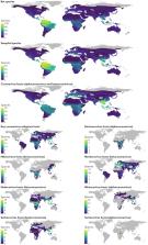
          In the past two decades, three coronaviruses with ancestral origins in bats have emerged and caused widespread outbreaks in humans, including severe acute respiratory syndrome coronavirus 2 (SARS-CoV-2). Since the first SARS epidemic in 2002–2003, the appreciation of bats as key hosts of zoonotic coronaviruses has advanced rapidly. More than 4,000 coronavirus sequences from 14 bat families have been identified, yet the true diversity of bat coronaviruses is probably much greater. Given that bats are the likely evolutionary source for several human coronaviruses, including strains that cause mild upper respiratory tract disease, their role in historic and future pandemics requires ongoing investigation. We review and integrate information on bat–coronavirus interactions at the molecular, tissue, host and population levels. We identify critical gaps in knowledge of bat coronaviruses, which relate to spillover and pandemic risk, including the pathways to zoonotic spillover, the infection dynamics within bat reservoir hosts, the role of prior adaptation in intermediate hosts for zoonotic transmission and the viral genotypes or traits that predict zoonotic capacity and pandemic potential. Filling these knowledge gaps may help prevent the next pandemic.

          Abstract

          Bats harbour a multitude of coronaviruses and owing to their diversity and wide distribution are prime reservoir hosts of emerging viruses. Ruiz-Aravena, McKee and colleagues analyse the currently available information on bat coronaviruses and discuss their role in recent and potential future spillovers.

          Related collections

          Most cited references173

          • Record: found
          • Abstract: found
          • Article: found
          Is Open Access

          A pneumonia outbreak associated with a new coronavirus of probable bat origin

          Since the outbreak of severe acute respiratory syndrome (SARS) 18 years ago, a large number of SARS-related coronaviruses (SARSr-CoVs) have been discovered in their natural reservoir host, bats 1–4 . Previous studies have shown that some bat SARSr-CoVs have the potential to infect humans 5–7 . Here we report the identification and characterization of a new coronavirus (2019-nCoV), which caused an epidemic of acute respiratory syndrome in humans in Wuhan, China. The epidemic, which started on 12 December 2019, had caused 2,794 laboratory-confirmed infections including 80 deaths by 26 January 2020. Full-length genome sequences were obtained from five patients at an early stage of the outbreak. The sequences are almost identical and share 79.6% sequence identity to SARS-CoV. Furthermore, we show that 2019-nCoV is 96% identical at the whole-genome level to a bat coronavirus. Pairwise protein sequence analysis of seven conserved non-structural proteins domains show that this virus belongs to the species of SARSr-CoV. In addition, 2019-nCoV virus isolated from the bronchoalveolar lavage fluid of a critically ill patient could be neutralized by sera from several patients. Notably, we confirmed that 2019-nCoV uses the same cell entry receptor—angiotensin converting enzyme II (ACE2)—as SARS-CoV.
            Bookmark
            • Record: found
            • Abstract: found
            • Article: found
            Is Open Access

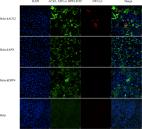
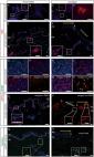
            Re-epithelialization and immune cell behaviour in an ex vivo human skin model

            A large body of literature is available on wound healing in humans. Nonetheless, a standardized ex vivo wound model without disruption of the dermal compartment has not been put forward with compelling justification. Here, we present a novel wound model based on application of negative pressure and its effects for epidermal regeneration and immune cell behaviour. Importantly, the basement membrane remained intact after blister roof removal and keratinocytes were absent in the wounded area. Upon six days of culture, the wound was covered with one to three-cell thick K14+Ki67+ keratinocyte layers, indicating that proliferation and migration were involved in wound closure. After eight to twelve days, a multi-layered epidermis was formed expressing epidermal differentiation markers (K10, filaggrin, DSG-1, CDSN). Investigations about immune cell-specific manners revealed more T cells in the blister roof epidermis compared to normal epidermis. We identified several cell populations in blister roof epidermis and suction blister fluid that are absent in normal epidermis which correlated with their decrease in the dermis, indicating a dermal efflux upon negative pressure. Together, our model recapitulates the main features of epithelial wound regeneration, and can be applied for testing wound healing therapies and investigating underlying mechanisms.
              Bookmark
              • Record: found
              • Abstract: found
              • Article: not found

              Tissue distribution of ACE2 protein, the functional receptor for SARS coronavirus. A first step in understanding SARS pathogenesis

              Abstract Severe acute respiratory syndrome (SARS) is an acute infectious disease that spreads mainly via the respiratory route. A distinct coronavirus (SARS‐CoV) has been identified as the aetiological agent of SARS. Recently, a metallopeptidase named angiotensin‐converting enzyme 2 (ACE2) has been identified as the functional receptor for SARS‐CoV. Although ACE2 mRNA is known to be present in virtually all organs, its protein expression is largely unknown. Since identifying the possible route of infection has major implications for understanding the pathogenesis and future treatment strategies for SARS, the present study investigated the localization of ACE2 protein in various human organs (oral and nasal mucosa, nasopharynx, lung, stomach, small intestine, colon, skin, lymph nodes, thymus, bone marrow, spleen, liver, kidney, and brain). The most remarkable finding was the surface expression of ACE2 protein on lung alveolar epithelial cells and enterocytes of the small intestine. Furthermore, ACE2 was present in arterial and venous endothelial cells and arterial smooth muscle cells in all organs studied. In conclusion, ACE2 is abundantly present in humans in the epithelia of the lung and small intestine, which might provide possible routes of entry for the SARS‐CoV. This epithelial expression, together with the presence of ACE2 in vascular endothelium, also provides a first step in understanding the pathogenesis of the main SARS disease manifestations. Copyright © 2004 Pathological Society of Great Britain and Ireland. Published by John Wiley & Sons, Ltd.
                Bookmark

                Author and article information

                Contributors
                raina.plowright@montana.edu
                Journal
                Nat Rev Microbiol
                Nat Rev Microbiol
                Nature Reviews. Microbiology
                Nature Publishing Group UK (London )
                1740-1526
                1740-1534
                19 November 2021
                : 1-16
                Affiliations
                [1 ]GRID grid.41891.35, ISNI 0000 0001 2156 6108, Department of Microbiology and Cell Biology, , Montana State University, ; Bozeman, MT USA
                [2 ]GRID grid.21107.35, ISNI 0000 0001 2171 9311, Department of Epidemiology, , Johns Hopkins Bloomberg School of Public Health, ; Baltimore, MD USA
                [3 ]GRID grid.19006.3e, ISNI 0000 0000 9632 6718, Department of Ecology and Evolutionary Biology, , University of California, Los Angeles, ; Los Angeles, CA USA
                [4 ]GRID grid.1022.1, ISNI 0000 0004 0437 5432, Centre for Planetary Health and Food Security, , Griffith University, ; Nathan, QLD Australia
                [5 ]GRID grid.5335.0, ISNI 0000000121885934, Department of Veterinary Medicine, , University of Cambridge, ; Cambridge, UK
                [6 ]GRID grid.419681.3, ISNI 0000 0001 2164 9667, National Institute of Allergy and Infectious Diseases, ; Hamilton, MT USA
                [7 ]GRID grid.5386.8, ISNI 000000041936877X, Department of Microbiology and Immunology, College of Veterinary Medicine, , Cornell University, ; Ithaca, NY USA
                [8 ]GRID grid.29857.31, ISNI 0000 0001 2097 4281, Department of Biology, Center for Infectious Disease Dynamics, , Pennsylvania State University, ; University Park, PA USA
                [9 ]GRID grid.170205.1, ISNI 0000 0004 1936 7822, Department of Ecology and Evolution, , University of Chicago, ; Chicago, IL USA
                [10 ]GRID grid.47894.36, ISNI 0000 0004 1936 8083, Department of Microbiology, Immunology, and Pathology, College of Veterinary Medicine and Biomedical Sciences, , Colorado State University, ; Fort Collins, CO USA
                [11 ]GRID grid.41891.35, ISNI 0000 0001 2156 6108, Department of Ecology, , Montana State University, ; Bozeman, MT USA
                Author information
                http://orcid.org/0000-0001-8463-7858
                http://orcid.org/0000-0002-7604-2296
                http://orcid.org/0000-0001-5380-5281
                http://orcid.org/0000-0003-1940-7794
                http://orcid.org/0000-0003-3538-3550
                http://orcid.org/0000-0001-9158-853X
                http://orcid.org/0000-0002-1292-7650
                http://orcid.org/0000-0002-1836-6655
                http://orcid.org/0000-0001-7941-502X
                http://orcid.org/0000-0002-2288-3196
                http://orcid.org/0000-0002-3338-6590
                Article
                652
                10.1038/s41579-021-00652-2
                8603903
                34799704
                4848bbc6-51cd-42ca-abc1-ed7ecf17b503
                © Springer Nature Limited 2021

                This article is made available via the PMC Open Access Subset for unrestricted research re-use and secondary analysis in any form or by any means with acknowledgement of the original source. These permissions are granted for the duration of the World Health Organization (WHO) declaration of COVID-19 as a global pandemic.

                History
                : 19 October 2021
                Categories
                Review Article

                sars-cov-2,ecology,infectious diseases,viral reservoirs
                sars-cov-2, ecology, infectious diseases, viral reservoirs

                Comments

                Comment on this article