1
views
0
recommends
+1 Recommend
0 collections
    0
    shares
      • Record: found
      • Abstract: found
      • Article: found
      Is Open Access

      Menopausal hormone therapy and the female brain: leveraging neuroimaging and prescription registry data from the UK Biobank cohort.

      Preprint
      research-article

      Read this article at

      Bookmark
          There is no author summary for this article yet. Authors can add summaries to their articles on ScienceOpen to make them more accessible to a non-specialist audience.

          Abstract

          Background and Objectives:

          Menopausal hormone therapy (MHT) is generally thought to be neuroprotective, yet results have been inconsistent. Here, we present a comprehensive study of MHT use and brain characteristics in middle- to older aged females from the UK Biobank, assessing detailed MHT data, APOE ε4 genotype, and tissue-specific gray (GM) and white matter (WM) brain age gap (BAG), as well as hippocampal and white matter hyperintensity (WMH) volumes.

          Methods:

          A total of 19,846 females with magnetic resonance imaging data were included (current-users = 1,153, 60.1 ± 6.8 years; past-users = 6,681, 67.5 ± 6.2 years; never-users = 12,012, mean age 61.6 ± 7.1 years). For a sub-sample (n = 538), MHT prescription data was extracted from primary care records. Brain measures were derived from T1-, T2- and diffusion-weighted images. We fitted regression models to test for associations between the brain measures and MHT variables including user status, age at initiation, dosage and duration, formulation, route of administration, and type (i.e., bioidentical vs synthetic), as well as active ingredient (e.g., estradiol hemihydrate). We further tested for differences in brain measures among MHT users with and without a history of hysterectomy ± bilateral oophorectomy and examined associations by APOE ε4 status.

          Results:

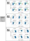
          We found significantly higher GM and WM BAG (i.e., older brain age relative to chronological age) as well as smaller left and right hippocampus volumes in current MHT users, not past users, compared to never-users. Effects were modest, with the largest effect size indicating a group difference of 0.77 years (~9 months) for GM BAG. Among MHT users, we found no significant associations between age at MHT initiation and brain measures. Longer duration of use and older age at last use post menopause was associated with higher GM and WM BAG, larger WMH volume, and smaller left and right hippocampal volumes. MHT users with a history of hysterectomy ± bilateral oophorectomy showed lower GM BAG relative to MHT users without such history. Although we found smaller hippocampus volumes in carriers of two APOE ε4 alleles compared to non-carriers, we found no interactions with MHT variables. In the sub-sample with prescription data, we found no significant associations between detailed MHT variables and brain measures after adjusting for multiple comparisons.

          Discussion:

          Our results indicate that population-level associations between MHT use, and female brain health might vary depending on duration of use and past surgical history. Future research is crucial to establish causality, dissect interactions between menopause-related neurological changes and MHT use, and determine individual-level implications to advance precision medicine in female health care.

          Related collections

          Most cited references87

          • Record: found
          • Abstract: not found
          • Article: not found

          Controlling the False Discovery Rate: A Practical and Powerful Approach to Multiple Testing

            Bookmark
            • Record: found
            • Abstract: found
            • Article: found
            Is Open Access

            The UK Biobank resource with deep phenotyping and genomic data

            The UK Biobank project is a prospective cohort study with deep genetic and phenotypic data collected on approximately 500,000 individuals from across the United Kingdom, aged between 40 and 69 at recruitment. The open resource is unique in its size and scope. A rich variety of phenotypic and health-related information is available on each participant, including biological measurements, lifestyle indicators, biomarkers in blood and urine, and imaging of the body and brain. Follow-up information is provided by linking health and medical records. Genome-wide genotype data have been collected on all participants, providing many opportunities for the discovery of new genetic associations and the genetic bases of complex traits. Here we describe the centralized analysis of the genetic data, including genotype quality, properties of population structure and relatedness of the genetic data, and efficient phasing and genotype imputation that increases the number of testable variants to around 96 million. Classical allelic variation at 11 human leukocyte antigen genes was imputed, resulting in the recovery of signals with known associations between human leukocyte antigen alleles and many diseases.
              Bookmark
              • Record: found
              • Abstract: found
              • Article: not found

              A multi-modal parcellation of human cerebral cortex

              Understanding the amazingly complex human cerebral cortex requires a map (or parcellation) of its major subdivisions, known as cortical areas. Making an accurate areal map has been a century-old objective in neuroscience. Using multi-modal magnetic resonance images from the Human Connectome Project (HCP) and an objective semi-automated neuroanatomical approach, we delineated 180 areas per hemisphere bounded by sharp changes in cortical architecture, function, connectivity, and/or topography in a precisely aligned group average of 210 healthy young adults. We characterized 97 new areas and 83 areas previously reported using post-mortem microscopy or other specialized study-specific approaches. To enable automated delineation and identification of these areas in new HCP subjects and in future studies, we trained a machine-learning classifier to recognize the multi-modal ‘fingerprint’ of each cortical area. This classifier detected the presence of 96.6% of the cortical areas in new subjects, replicated the group parcellation, and could correctly locate areas in individuals with atypical parcellations. The freely available parcellation and classifier will enable substantially improved neuroanatomical precision for studies of the structural and functional organization of human cerebral cortex and its variation across individuals and in development, aging, and disease.
                Bookmark

                Author and article information

                Journal
                medRxiv
                MEDRXIV
                medRxiv
                Cold Spring Harbor Laboratory
                12 April 2024
                : 2024.04.08.24305450
                Affiliations
                [1 ]Department of Psychiatric Research, Diakonhjemmet Hospital, Oslo, Norway
                [2 ]Department of Psychiatry, University of Toronto, Toronto, ON, Canada
                [3 ]Centre for Addiction and Mental Health, Toronto, ON, Canada
                [4 ]Psychological and Brain Sciences, University of California Santa Barbara, CA, USA
                [5 ]Department of Psychology, University of Oslo, Oslo, Norway
                [6 ]Centre for Precision Psychiatry, Division of Mental Health and Addiction, Oslo University Hospital, Oslo, Norway
                [7 ]Department of Clinical Neurosciences, Lausanne University Hospital (CHUV) and University of Lausanne, Lausanne, Switzerland
                [8 ]Department of Psychiatry, University of Oxford, Oxford, UK
                Author notes
                [* ] Corresponding author: Claudia Barth, PhD, claudia.barth@ 123456medisin.uio.no
                Author information
                http://orcid.org/0000-0001-6544-0945
                http://orcid.org/0000-0003-2874-9972
                http://orcid.org/0000-0003-0001-5096
                http://orcid.org/0000-0001-8644-956X
                http://orcid.org/0000-0002-5150-6656
                Article
                10.1101/2024.04.08.24305450
                11030497
                38645009
                461b507b-a710-4f02-ab80-d1a27b01630b

                This work is licensed under a Creative Commons Attribution-NonCommercial-NoDerivatives 4.0 International License, which allows reusers to copy and distribute the material in any medium or format in unadapted form only, for noncommercial purposes only, and only so long as attribution is given to the creator.

                History
                Funding
                The authors received funding from the Research Council of Norway (LTW: 223273, 249795, 273345, 298646, 300768), the South-Eastern Norway Regional Health Authority (CB: 2023037, 2022103; LTW: 2018076, 2019101), the European Research Council under the European Union’s Horizon 2020 research and innovation programme (LTW: 802998), the Swiss National Science Foundation (AMGdL: PZ00P3_193658), the Canadian Institutes for Health Research (LAMG: PJT-173554), the Treliving Family Chair in Women’s Mental Health at the Centre for Addiction and Mental Health (LAMG), womenmind at the Centre for Addiction and Mental Health (LAMG, BHL), the Ann S. Bowers Women’s Brain Health Initiative (EGJ), and the National Institutes of Health (EGJ: AG063843).
                Categories
                Article

                Comments

                Comment on this article