31
views
0
recommends
+1 Recommend
0 collections
    3
    shares
      • Record: found
      • Abstract: found
      • Article: found
      Is Open Access

      Glaucoma and Corneal Transplant Procedures

      review-article

      Read this article at

      Bookmark
          There is no author summary for this article yet. Authors can add summaries to their articles on ScienceOpen to make them more accessible to a non-specialist audience.

          Abstract

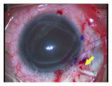
          Glaucoma after corneal transplantation is a leading cause of ocular morbidity after penetrating keratoplasty. The incidence reported is highly variable and a number of etiologic factors have been identified. A number of treatment options are available; surgical intervention for IOP control is associated with a high incidence of graft failure. IOP elevation is less frequently seen following deep anterior lamellar keratoplasty. Descemet's striping-automated endothelial keratoplasty is also associated with postprocedure intraocular pressure elevation and secondary glaucoma and presents unique surgical challenges in patients with preexisting glaucoma surgeries. Glaucoma exists in up to three-quarters of patients who undergo keratoprosthesis surgery and the management if often challenging. The aim of this paper is to highlight the incidence, etiology, and management of glaucoma following different corneal transplant procedures. It also focuses on the challenges in the diagnosis of glaucoma and intraocular pressure monitoring in this group of patients.

          Related collections

          Most cited references87

          • Record: found
          • Abstract: found
          • Article: not found

          Descemet's stripping endothelial keratoplasty: safety and outcomes: a report by the American Academy of Ophthalmology.

          To review the published literature on safety and outcomes of Descemet's stripping endothelial keratoplasty (DSEK) for the surgical treatment of endothelial diseases of the cornea. Peer-reviewed literature searches were conducted in PubMed and the Cochrane Library with the most recent search in February 2009. The searches yielded 2118 citations in English-language journals. The abstracts of these articles were reviewed and 131 articles were selected for possible clinical relevance, of which 34 were determined to be relevant to the assessment objectives. The most common complications from DSEK among reviewed reports included posterior graft dislocations (mean, 14%; range, 0%-82%), followed by endothelial graft rejection (mean, 10%; range, 0%-45%), primary graft failure (mean, 5%; range, 0%-29%), and iatrogenic glaucoma (mean, 3%; range, 0%-15%). Average endothelial cell loss as measured by specular microscopy ranged from 25% to 54%, with an average cell loss of 37% at 6 months, and from 24% to 61%, with an average cell loss of 42% at 12 months. The average best-corrected Snellen visual acuity (mean, 9 months; range, 3-21 months) ranged from 20/34 to 20/66. A review of postoperative refractive results found induced hyperopia ranging from 0.7 to 1.5 diopters (D; mean, 1.1 D), with minimal induced astigmatism ranging from -0.4 to 0.6 D and a mean refractive shift of 0.11 D. A review of graft survival found that clear grafts at 1 year ranged from 55% to 100% (mean, 94%). The evidence reviewed is supportive of DSEK being a safe and effective treatment for endothelial diseases of the cornea. In terms of surgical risks, complication rates, graft survival (clarity), visual acuity, and endothelial cell loss, DSEK appears similar to penetrating keratoplasty (PK). It seems to be superior to PK in terms of earlier visual recovery, refractive stability, postoperative refractive outcomes, wound and suture-related complications, and intraoperative and late suprachoroidal hemorrhage risk. The most common complications of DSEK do not appear to be detrimental to the ultimate vision recovery in most cases. Long-term endothelial cell survival and the risk of late endothelial rejection are beyond the scope of this assessment. Proprietary or commercial disclosure may be found after the references.
            Bookmark
            • Record: found
            • Abstract: found
            • Article: not found

            Corticosteroid-induced glaucoma: a review of the literature.

            The intraocular pressure rise that can complicate the use of topical or systemic corticosteroid has been recognised for 50 years. More recently, following isolation of the myocilin gene (previously known as the trabecular meshwork inducible glucocorticoid response or TIGR gene), there has been renewed interest in this steroid-responsive phenomenon. Furthermore, the currently fashionable use of injectable intraocular steroids in the management of clinically significant subretinal fluid and macular oedema has resulted in an increased incidence. Animal studies, cell biology, molecular biology, and an improved knowledge of genetics have provided a better understanding of the mechanisms behind the response. The purpose of this review is to describe the risk factors for developing corticosteroid-induced glaucoma, to discuss the underlying mechanisms and genetics of the condition and to present management options.
              Bookmark
              • Record: found
              • Abstract: found
              • Article: not found

              Distribution of central corneal thickness and its association with intraocular pressure: The Rotterdam Study.

              To perform a cross-sectional study on the distribution of central corneal thickness and its association with intraocular pressure in an elderly population. We measured central corneal thickness and intraocular pressure in 395 subjects (352 control subjects, 13 patients with ocular hypertension, and 30 patients with primary open-angle glaucoma) aged 55 years or more. Mean central corneal thickness in the 352 control subjects was 537.4 microm (95% confidence interval [CI], 533.8 to 540.9 microm; range, 427 to 620 microm), with a maximal difference between eyes of 42 microm. There were no differences between sexes and no significant association with age. Linear regression analysis showed an increase of 0.19 mm Hg in intraocular pressure with each 10-microm increase in central corneal thickness (95% CI, 0.09 to 0.28 mm Hg). This association was similar in both eyes and in both sexes. The 13 patients with ocular hypertension had corneas a mean of 16.0 microm thicker (95% CI, -2.6 to +34.6 microm) compared with control subjects (P = .093); the 30 patients with primary open-angle glaucoma had corneas a mean of 21.5 microm thinner (95% CI, 8.8 to 34.1 microm) compared with control subjects (P = .001). Mean central corneal thickness was similar to that found in clinical studies, was slightly higher in patients with ocular hypertension, and was significantly lower in patients with primary open-angle glaucoma. Intraocular pressure was positively related with central corneal thickness. Central corneal thickness may influence the division between normal and increased intraocular pressure at a simple cutoff point of 21 mm Hg.
                Bookmark

                Author and article information

                Journal
                J Ophthalmol
                JOP
                Journal of Ophthalmology
                Hindawi Publishing Corporation
                2090-004X
                2090-0058
                2012
                18 January 2012
                : 2012
                : 576394
                Affiliations
                1Department of Ophthalmology, Ministry of Health, Manama 12, Bahrain
                2Division of Anterior Segment, King Khaled Eye Specialists Hospital, Riyadh 11462, Saudi Arabia
                3Wilmer Eye Institute, Johns Hopkins University School of Medicine, Baltimore, MD 21287, USA
                Author notes
                *Deepak P. Edward: dedwar39@ 123456jhmi.edu

                Academic Editor: G. L. Spaeth

                Article
                10.1155/2012/576394
                3270571
                22315661
                40716171-a5e0-42ad-b5ca-c27966177efc
                Copyright © 2012 Ammar M. Al-Mahmood et al.

                This is an open access article distributed under the Creative Commons Attribution License, which permits unrestricted use, distribution, and reproduction in any medium, provided the original work is properly cited.

                History
                : 1 August 2011
                : 10 October 2011
                Categories
                Review Article

                Ophthalmology & Optometry
                Ophthalmology & Optometry

                Comments

                Comment on this article