19
views
0
recommends
+1 Recommend
0 collections
    0
    shares
      • Record: found
      • Abstract: found
      • Article: found
      Is Open Access

      Effect of Zishen Jiangtang Pill, a Chinese Herbal Product, on Rats with Diabetic Osteoporosis

      research-article

      Read this article at

      Bookmark
          There is no author summary for this article yet. Authors can add summaries to their articles on ScienceOpen to make them more accessible to a non-specialist audience.

          Abstract

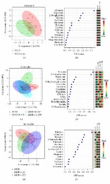
          Diabetic osteoporosis (DO) is a complication of diabetes. Zishen Jiangtang Pill (ZJP) is a Chinese herbal product which has been used in clinic to maintain blood glucose level and bone density for decades. However, the evidence about its mechanism on diabetes and osteoporosis is still unknown. The aim of this study is to investigate therapeutic effect of ZJP on DO in streptozotocin- (STZ-) induced rats. Rats were randomly assigned to 4 groups: one control group (CON), one model group (MOD), and two ZJP treatment groups (1.5 and 3.0 g/kg/d). All rats were treated for 8 weeks. Results showed that ZJP decreased the blood glucose level during OGTT and prevented the changes of FBG and Fins. Similarly, ZJP inhibited the changes of BCa, P, TRACP-5b, CTX-1, BALP, and BGP and the reduction of BMD. In parallel, 1H-NMR metabolomic studies showed that ZJP significantly altered the metabolic fingerprints of blood and urine level. These findings suggest that ZJP can effectively improve glucose metabolism, abnormal bone metabolism, and metabolic disorders in DO rats, which may be a useful alternative medicine for DO therapy.

          Related collections

          Most cited references29

          • Record: found
          • Abstract: found
          • Article: not found

          Osteoporosis: now and the future.

          Osteoporosis is a common disease characterised by a systemic impairment of bone mass and microarchitecture that results in fragility fractures. With an ageing population, the medical and socioeconomic effect of osteoporosis, particularly postmenopausal osteoporosis, will increase further. A detailed knowledge of bone biology with molecular insights into the communication between bone-forming osteoblasts and bone-resorbing osteoclasts and the orchestrating signalling network has led to the identification of novel therapeutic targets. Novel treatment strategies have been developed that aim to inhibit excessive bone resorption and increase bone formation. The most promising novel treatments include: denosumab, a monoclonal antibody for receptor activator of NF-κB ligand, a key osteoclast cytokine; odanacatib, a specific inhibitor of the osteoclast protease cathepsin K; and antibodies against the proteins sclerostin and dickkopf-1, two endogenous inhibitors of bone formation. This overview discusses these novel therapies and explains their underlying physiology. Copyright © 2011 Elsevier Ltd. All rights reserved.
            Bookmark
            • Record: found
            • Abstract: found
            • Article: not found

            Bone marrow, cytokines, and bone remodeling. Emerging insights into the pathophysiology of osteoporosis.

            Both osteoblasts and osteoclasts are derived from progenitors that reside in the bone marrow; osteoblasts belong to the mesenchymal lineage of the marrow stroma, and osteoclasts to the hematopoietic lineage. The development of osteoclasts from their progenitors is dependent on stromal-osteoblastic cells, which are a major source of cytokines that are critical in osteoclastogenesis, such as interleukin-6 and interleukin-11. The production of interleukin-6 by stromal osteoblastic cells, as well as the responsiveness of bone marrow cells to cytokines such as interleukin-6 and interleukin-11, is regulated by sex steroids. When gonadal function is lost, the formation of osteoclasts as well as osteoblasts increases in the marrow, both changes apparently mediated by an increase in the production of interleukin-6 and perhaps by an increase in the responsiveness of bone marrow progenitor cells not only to interleukin-6 but also to other cytokines with osteoclastogenic and osteoblastogenic properties. The cellular activity of the bone marrow is also altered by the process of aging. Specifically, senescence may decrease the ability of the marrow to form osteoblast precursors. The association between the dysregulation of osteoclast or osteoblast development in the marrow and the disruption of the balance between bone resorption and bone formation, resulting in the loss of bone, leads to the following notion. Like homeostasis of other regenerating tissues, homeostasis of bone depends on the orderly replenishment of its cellular constituents. Excessive osteoclastogenesis and inadequate osteoblastogenesis are responsible for the mismatch between the formation and resorption of bone in postmenopausal and age-related osteopenia. The recognition that changes in the numbers of bone cells, rather than changes in the activity of individual cells, form the pathogenetic basis of osteoporosis is a major advance in understanding the mechanism of this disease.
              Bookmark
              • Record: found
              • Abstract: found
              • Article: found
              Is Open Access

              High Bone Mineral Density and Fracture Risk in Type 2 Diabetes as Skeletal Complications of Inadequate Glucose Control

              OBJECTIVE Individuals with type 2 diabetes have increased fracture risk despite higher bone mineral density (BMD). Our aim was to examine the influence of glucose control on skeletal complications. RESEARCH DESIGN AND METHODS Data of 4,135 participants of the Rotterdam Study, a prospective population-based cohort, were available (mean follow-up 12.2 years). At baseline, 420 participants with type 2 diabetes were classified by glucose control (according to HbA1c calculated from fructosamine), resulting in three comparison groups: adequately controlled diabetes (ACD; n = 203; HbA1c <7.5%), inadequately controlled diabetes (ICD; n = 217; HbA1c ≥7.5%), and no diabetes (n = 3,715). Models adjusted for sex, age, height, and weight (and femoral neck BMD) were used to test for differences in bone parameters and fracture risk (hazard ratio [HR] [95% CI]). RESULTS The ICD group had 1.1–5.6% higher BMD, 4.6–5.6% thicker cortices, and −1.2 to −1.8% narrower femoral necks than ACD and ND, respectively. Participants with ICD had 47–62% higher fracture risk than individuals without diabetes (HR 1.47 [1.12–1.92]) and ACD (1.62 [1.09–2.40]), whereas those with ACD had a risk similar to those without diabetes (0.91 [0.67–1.23]). CONCLUSIONS Poor glycemic control in type 2 diabetes is associated with fracture risk, high BMD, and thicker femoral cortices in narrower bones. We postulate that fragility in apparently “strong” bones in ICD can result from microcrack accumulation and/or cortical porosity, reflecting impaired bone repair.
                Bookmark

                Author and article information

                Contributors
                Journal
                Evid Based Complement Alternat Med
                Evid Based Complement Alternat Med
                ECAM
                Evidence-based Complementary and Alternative Medicine : eCAM
                Hindawi
                1741-427X
                1741-4288
                2018
                12 February 2018
                : 2018
                : 7201914
                Affiliations
                Shenzhen Traditional Chinese Medicine Hospital, Guangzhou University of Chinese Medicine, Shenzhen 518033, China
                Author notes

                Academic Editor: Vicky P. Chen

                Author information
                http://orcid.org/0000-0002-4888-1025
                http://orcid.org/0000-0002-7134-8173
                http://orcid.org/0000-0003-1327-1902
                http://orcid.org/0000-0002-5502-7820
                http://orcid.org/0000-0001-7511-6049
                http://orcid.org/0000-0003-1315-7739
                http://orcid.org/0000-0003-1892-9355
                http://orcid.org/0000-0002-7892-1112
                http://orcid.org/0000-0002-0417-2243
                Article
                10.1155/2018/7201914
                5833190
                3d58b684-9675-49eb-b119-77b9b1a05db1
                Copyright © 2018 Huilin Li et al.

                This is an open access article distributed under the Creative Commons Attribution License, which permits unrestricted use, distribution, and reproduction in any medium, provided the original work is properly cited.

                History
                : 31 August 2017
                : 13 November 2017
                : 24 December 2017
                Funding
                Funded by: Shenzhen Scientific Research Fund
                Award ID: JCYJ20140408153331810
                Award ID: ZDSYS201606081515458
                Categories
                Research Article

                Complementary & Alternative medicine
                Complementary & Alternative medicine

                Comments

                Comment on this article