55
views
0
recommends
+1 Recommend
0 collections
    0
    shares
      • Record: found
      • Abstract: found
      • Article: found
      Is Open Access

      Early Visual Processing is Affected by Clinical Subtype in Patients with Unilateral Spatial Neglect: A Magnetoencephalography Study

      research-article

      Read this article at

      Bookmark
          There is no author summary for this article yet. Authors can add summaries to their articles on ScienceOpen to make them more accessible to a non-specialist audience.

          Abstract

          Objective: To determine whether visual evoked magnetic fields (VEFs) elicited by right and left hemifield stimulation differ in patients with unilateral spatial neglect (USN) that results from cerebrovascular accident.

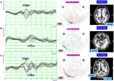
          Methods: Pattern-reversal stimulation of the right and left hemifield was performed in three patients with left USN. Magnetoencephalography (MEG) was recorded using a 160-channel system, and VEFs were quantified in the 400 ms after each stimulus. The presence or absence of VEF components at around 100 ms (P100m component) and 145 ms (N145m component) after stimulus onset was determined. The source of the VEF was determined using a single equivalent current dipole model for spherical volume conduction. All patients were evaluated using the behavioral inattention test (BIT).

          Results: In response to right hemifield stimulation, the P100m and N145m components of the VEF were evident in all three patients. In response to left hemifield stimulation, both components were evident in Patient 3, whereas only the P100m component was evident in Patient 1 and only the N145m component was evident in Patient 2. Patient 1 exhibited impairments on the line bisection and cancelation tasks of the BIT, Patient 2 exhibited impairments on the copying, drawing and cancelation tasks of the BIT, and Patient 3 exhibited impairments on the cancelation task of the BIT.

          Conclusion: These results demonstrate that early VEFs are disrupted in patients with USN and support the concept that deficits in visual processing differ according to the clinical subtype of USN and the lesion location. This study also demonstrates the feasibility of using MEG to explore subtypes of neglect.

          Related collections

          Most cited references36

          • Record: found
          • Abstract: found
          • Article: not found

          Spatial attention and neglect: parietal, frontal and cingulate contributions to the mental representation and attentional targeting of salient extrapersonal events.

          The syndrome of contralesional neglect reflects a lateralized disruption of spatial attention. In the human, the left hemisphere shifts attention predominantly in the contralateral hemispace and in a contraversive direction whereas the right hemisphere distributes attention more evenly, in both hemispaces and both directions. As a consequence of this asymmetry, severe contralesional neglect occurs almost exclusively after right hemisphere lesions. Patients with left neglect experience a loss of salience in the mental representation and conscious perception of the left side and display a reluctance to direct orientating and exploratory behaviours to the left. Neglect is distributed according to egocentric, allocentric, world-centred, and object-centred frames of reference. Neglected events can continue to exert an implicit influence on behaviour, indicating that the attentional filtering occurs at the level of an internalized representation rather than at the level of peripheral sensory input. The unilateral neglect syndrome is caused by a dysfunction of a large-scale neurocognitive network, the cortical epicentres of which are located in posterior parietal cortex, the frontal eye fields, and the cingulate gyrus. This network coordinates all aspects of spatial attention, regardless of the modality of input or output. It helps to compile a mental representation of extrapersonal events in terms of their motivational salience, and to generate 'kinetic strategies' so that the attentional focus can shift from one target to another.
            Bookmark
            • Record: found
            • Abstract: found
            • Article: not found

            Magnetoencephalography: evidence of magnetic fields produced by alpha-rhythm currents.

            D. Cohen (1968)
            Weak alternating magnetic fields outside the human scalp, produced by alpha-rhythm currents, are demonstrated. Subject ard magnetic detector were housed in a multilayer magnetically shielded chamber. Background magnetic noise was reduced by signal-averaging. The fields near the scalp are about 1 x 10(-9) gauss (peak to peak). A course distribution shows left-right symmetry for the particular averaging technique used here.
              Bookmark
              • Record: found
              • Abstract: found
              • Article: not found

              Direct evidence for a parietal-frontal pathway subserving spatial awareness in humans.

              Intraoperative electrical stimulation, which temporarily inactivates restricted regions during brain surgery, can map cognitive functions in humans with spatiotemporal resolution unmatched by other methods. Using this technique, we found that stimulation of the right inferior parietal lobule or the caudal superior temporal gyrus, but not of its rostral portion, determined rightward deviations on line bisection. However, the strongest shifts occurred with subcortical stimulation. Fiber tracking identified the stimulated site as a section of the superior occipitofrontal fasciculus, a poorly known parietal-frontal pathway. These findings suggest that parietal-frontal communication is necessary for the symmetrical processing of the visual scene.
                Bookmark

                Author and article information

                Journal
                Front Hum Neurosci
                Front Hum Neurosci
                Front. Hum. Neurosci.
                Frontiers in Human Neuroscience
                Frontiers Media S.A.
                1662-5161
                31 July 2013
                2013
                : 7
                : 432
                Affiliations
                [1] 1Department of Rehabilitation Medicine, Keio University School of Medicine , Tokyo, Japan
                [2] 2ImpAct Team, INSERM U1028, CNRS UMR5292, Lyon Neuroscience Research Center , Bron, France
                [3] 3National Sanatorium Tama Zenshoen , Tokyo, Japan
                [4] 4Makuhari Ohde Eye Clinic , Chiba, Japan
                [5] 5Department of Ophthalmology, Keio University School of Medicine , Tokyo, Japan
                Author notes

                Edited by: Tanja Nijboer, Utrecht University, Netherlands

                Reviewed by: Shozo Tobimatsu, Kyushu University, Japan; Maarten Van Der Smagt, Utrecht University, Netherlands

                *Correspondence: Tetsuya Tsuji, Department of Rehabilitation Medicine, Keio University School of Medicine, 35 Shinanomachi, Shinjuku-ku, Tokyo 160-8582, Japan e-mail: cxa01423@ 123456nifty.com
                Article
                10.3389/fnhum.2013.00432
                3728490
                23914171
                35a41229-1d54-4bdb-81b6-e256d3520d3a
                Copyright © 2013 Mizuno, Tsuji, Rossetti, Pisella, Ohde and Liu.

                This is an open-access article distributed under the terms of the Creative Commons Attribution License (CC BY). The use, distribution or reproduction in other forums is permitted, provided the original author(s) or licensor are credited and that the original publication in this journal is cited, in accordance with accepted academic practice. No use, distribution or reproduction is permitted which does not comply with these terms.

                History
                : 25 January 2013
                : 15 July 2013
                Page count
                Figures: 3, Tables: 3, Equations: 0, References: 39, Pages: 8, Words: 6231
                Categories
                Neuroscience
                Original Research

                Neurosciences
                visual evoked magnetic field,pattern-reversal stimulation,attention network,diagnosis of unilateral spatial neglect,neglect subtypes,visual attention networks,viewer-centered neglect,stimulus-centered neglect

                Comments

                Comment on this article