3
views
0
recommends
+1 Recommend
0 collections
    0
    shares
      • Record: found
      • Abstract: found
      • Article: found
      Is Open Access

      Multimodality imaging in lobular breast cancer: Differences in mammography, ultrasound, and MRI in the assessment of local tumor extent and correlation with molecular characteristics

      research-article

      Read this article at

      Bookmark
          There is no author summary for this article yet. Authors can add summaries to their articles on ScienceOpen to make them more accessible to a non-specialist audience.

          Abstract

          Introduction

          Invasive lobular breast cancer (ILC) is a diagnostic challenge due to the diversity of morphological features. The objective of the study was to investigate the presentation and local extent of ILC using various imaging techniques and to assess the correlation between imaging and molecular profile.

          Materials and methods

          We reviewed 162 consecutive patients with ILC found on vacuum-assisted biopsy, who underwent evaluation of the lesion morphology and extent using ultrasound (US), mammography (MMG), and magnetic resonance imaging (MRI). Radiographic features were compared with ILC intrinsic subtype based on the expression of Ki-67 and estrogen, progesterone, and HER2 receptors.

          Results

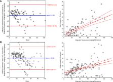
          A total of 113 mass lesions and 49 non-mass enhancements (NMEs) were found in MRI. Masses were typically irregular and spiculated, showing heterogeneous contrast enhancement, diffusion restriction, and type III enhancement curve. NMEs presented mainly as the area of focal or multiregional distribution with heterogeneous or clumped contrast enhancement, diffusion restriction, and type III enhancement curve. Lesion extent significantly varied between MRI and MMG/ultrasonography (USG) (P < 0.001) but did not differ between MGF and ultrasonography (USG). The larger the ILC, the higher the disproportion when lesion extent in MRI was compared with MMG (P < 0.001) and ultrasonography (USG) (P < 0.001). In the study group, there were 97 cases of luminal A subtype (59.9%), 54 cases of luminal B HER2− (33.3%), nine cases of luminal B HER2+ (5.5%), and two cases of triple negative (1.2%). The HER2 type was not found in the study group. We did not observe any significant correlation between molecular profile and imaging.

          Conclusion

          MRI is the most effective technique for the assessment of ILC local extent, which is important for optimal treatment planning. Further studies are needed to investigate if the intrinsic subtype of ILC can be predicted by imaging features on MRI.

          Related collections

          Most cited references48

          • Record: found
          • Abstract: found
          • Article: not found

          The HER-2 receptor and breast cancer: ten years of targeted anti-HER-2 therapy and personalized medicine.

          The human epidermal growth factor receptor (HER-2) oncogene encodes a transmembrane tyrosine kinase receptor that has evolved as a major classifier of invasive breast cancer and target of therapy for the disease. The validation of the general prognostic significance of HER-2 gene amplification and protein overexpression in the absence of anti-HER-2 targeted therapy is discussed in a study of 107 published studies involving 39,730 patients, which produced an overall HER-2-positive rate of 22.2% and a mean relative risk for overall survival (OS) of 2.74. The issue of HER-2 status in primary versus metastatic breast cancer is considered along with a section on the features of metastatic HER-2-positive disease. The major marketed slide-based HER-2 testing approaches, immunohistochemistry, fluorescence in situ hybridization, and chromogenic in situ hybridization, are presented and contrasted in detail against the background of the published American Society of Clinical Oncology-College of American Pathologists guidelines for HER-2 testing. Testing issues, such as the impact of chromosome 17 polysomy and local versus central HER-2 testing, are also discussed. Emerging novel HER-2 testing techniques, including mRNA-based testing by real-time polymerase chain reaction and DNA microarray methods, HER-2 receptor dimerization, phosphorylated HER-2 receptors, and HER-2 status in circulating tumor cells, are also considered. A series of biomarkers potentially associated with resistance to trastuzumab is discussed with emphasis on the phosphatase and tensin homologue deleted on chromosome ten/Akt and insulin-like growth factor receptor pathways. The efficacy results for the more recently approved small molecule HER-1/HER-2 kinase inhibitor lapatinib are also presented along with a more limited review of markers of resistance for this agent. Additional topics in this section include combinations of both anti-HER-2 targeted therapies together as well as with novel agents including bevacizumab, everolimus, and tenespimycin. A series of novel HER-2-targeting agents is also presented, including pertuzumab, ertumaxomab, HER-2 vaccines, and recently discovered tyrosine kinase inhibitors. Biomarkers predictive of HER-2 targeted therapy toxicity are included, and the review concludes with a consideration of HER-2 status in the prediction of response to non-HER-2 targeted treatments including hormonal therapy, anthracyclines, and taxanes.
            Bookmark
            • Record: found
            • Abstract: found
            • Article: not found

            Diagnostic accuracy of mammography, clinical examination, US, and MR imaging in preoperative assessment of breast cancer.

            To prospectively assess accuracy of mammography, clinical examination, ultrasonography (US), and magnetic resonance (MR) imaging in preoperative assessment of local extent of breast cancer. Institutional review board approval and informed patient consent were obtained. Results of bilateral mammography, US, and contrast-enhanced MR imaging were analyzed from 111 consecutive women with known or suspected invasive breast cancer. Results were correlated with histopathologic findings. Analysis included 177 malignant foci in 121 cancerous breasts, of which 89 (50%) foci were palpable. Median size of 139 invasive foci was 18 mm (range, 2-107 mm). Mammographic sensitivity decreased from 100% in fatty breasts to 45% in extremely dense breasts. Mammographic sensitivity was highest for invasive ductal carcinoma (IDC) in 89 of 110 (81%) cases versus 10 of 29 (34%) cases of invasive lobular carcinoma (ILC) (P < .001) and 21 of 38 (55%) cases of ductal carcinoma in situ (DCIS) (P < .01). US showed higher sensitivity than did mammography for IDC, depicting 104 of 110 (94%) cases, and for ILC, depicting 25 of 29 (86%) cases (P < .01 for each). US showed higher sensitivity for invasive cancer than DCIS (18 of 38 [47%], P < .001). MR showed higher sensitivity than did mammography for all tumor types (P < .01) and higher sensitivity than did US for DCIS (P < .001), depicting 105 of 110 (95%) cases of IDC, 28 of 29 (96%) cases of ILC, and 34 of 38 (89%) cases of DCIS. In anticipation of conservation or no surgery after mammography and clinical examination in 96 breasts, additional tumor (which altered surgical approach) was present in 30. Additional tumor was depicted in 17 of 96 (18%) breasts at US and in 29 of 96 (30%) at MR, though extent was now overestimated in 12 of 96 (12%) at US and 20 of 96 (21%) at MR imaging. After combined mammography, clinical examination, and US, MR depicted additional tumor in another 12 of 96 (12%) breasts and led to overestimation of extent in another six (6%); US showed no detection benefit after MR imaging. Bilateral cancer was present in 10 of 111 (9%) patients; contralateral tumor was depicted mammographically in six and with both US and MR in an additional three. One contralateral cancer was demonstrated only clinically. In nonfatty breasts, US and MR imaging were more sensitive than mammography for invasive cancer, but both MR imaging and US involved risk of overestimation of tumor extent. Combined mammography, clinical examination, and MR imaging were more sensitive than any other individual test or combination of tests. (c) RSNA, 2004.
              Bookmark
              • Record: found
              • Abstract: found
              • Article: not found

              Infiltrating lobular carcinoma of the breast: tumor characteristics and clinical outcome

              Introduction Invasive lobular carcinoma (ILC) comprises approximately 10% of breast cancers and appears to have a distinct biology. Because it is less common than infiltrating ductal carcinoma (IDC), few data have been reported that address the biologic features of ILC in the context of their clinical outcome. In the present study we undertook an extensive comparison of ILC and IDC using a large database to provide a more complete and reliable assessment of their biologic phenotypes and clinical behaviors. Methods The clinical and biological features of 4140 patients with ILC were compared with those of 45,169 patients with IDC (not otherwise specified). The median follow-up period was 87 months. Results In comparison with IDC, ILC was significantly more likely to occur in older patients, to be larger in size, to be estrogen and progesterone receptor positive, to have lower S-phase fraction, to be diploid, and to be HER-2, p53, and epidermal growth factor receptor negative. It was more common for ILC than for IDC to metastasize to the gastrointestinal tract and ovary. The incidence of contralateral breast cancer was higher for ILC patients than for IDC patients (20.9% versus 11.2%; P < 0.0001). Breast preservation was modestly less frequent in ILC patients than in IDC patients. The 5-year disease-free survival was 85.7% for ILC and 83.5% for IDC (P = 0.13). The 5-year overall survival was 85.6% for ILC and 84.1% for IDC (P = 0.64). Conclusion Despite the fact that the biologic phenotype of ILC is quite favorable, these patients do not have better clinical outcomes than do patients with IDC. At present, management decisions should be based on individual patient and tumor biologic characteristics, and not on lobular histology.
                Bookmark

                Author and article information

                Contributors
                Journal
                Front Oncol
                Front Oncol
                Front. Oncol.
                Frontiers in Oncology
                Frontiers Media S.A.
                2234-943X
                22 August 2022
                2022
                : 12
                : 855519
                Affiliations
                [1] 1 Breast Unit, Department of Breast Imaging, Lower Silesian Oncology, Pulmonology and Hematology Center , Wroclaw, Poland
                [2] 2 Burn and Plastic Surgery Department, Ludwik Rydygier Memorial Specialized Hospital in Krakow , Krakow, Poland
                [3] 3 Department of Anatomy, Jagiellonian University Medical College , Krakow, Poland
                [4] 4 Breast Unit, Department of Breast Surgery, Lower Silesian Oncology, Pulmonology and Hematology Center , Wroclaw, Poland
                [5] 5 Department of Oncology, Faculty of Medicine, Wroclaw Medical University , Wroclaw, Poland
                Author notes

                Edited by: Marie-France Penet, Johns Hopkins Medicine, United States

                Reviewed by: Durgesh K. Dwivedi, King George’s Medical University, India; William Ian Duncombe Rae, The University of Sydney, Australia

                *Correspondence: Michał Lis, lis.michal.md@ 123456gmail.com

                †These authors have contributed equally to this work and share first authorship

                ‡These authors have contributed equally to this work and share last authorship

                This article was submitted to Breast Cancer, a section of the journal Frontiers in Oncology

                Article
                10.3389/fonc.2022.855519
                9441946
                31de88e1-ee55-46a6-98f3-d9b1725b52b6
                Copyright © 2022 Dołęga-Kozierowski, Lis, Marszalska-Jacak, Koziej, Celer, Bandyk, Kasprzak, Szynglarewicz and Matkowski

                This is an open-access article distributed under the terms of the Creative Commons Attribution License (CC BY). The use, distribution or reproduction in other forums is permitted, provided the original author(s) and the copyright owner(s) are credited and that the original publication in this journal is cited, in accordance with accepted academic practice. No use, distribution or reproduction is permitted which does not comply with these terms.

                History
                : 15 January 2022
                : 11 July 2022
                Page count
                Figures: 5, Tables: 4, Equations: 0, References: 48, Pages: 14, Words: 5771
                Funding
                Funded by: Uniwersytet Medyczny im. Piastów Slaskich we Wroclawiu , doi 10.13039/501100009687;
                Categories
                Oncology
                Original Research

                Oncology & Radiotherapy
                breast cancer,invasive lobular carcinoma (ilc),invasive lobular breast cancer,magnetic resonance imaging,multimodality imaging,lobular breast cancer

                Comments

                Comment on this article