65
views
0
recommends
+1 Recommend
2 collections
    0
    shares
      • Record: found
      • Abstract: found
      • Article: not found

      Evaluation of the mRNA-1273 Vaccine against SARS-CoV-2 in Nonhuman Primates

      research-article
      , Ph.D., , M.S., , Ph.D., , Ph.D., , Ph.D., , B.S., , Ph.D., , M.S., , M.B., , M.S., , M.S., , Ph.D., , Ph.D., , M.S., , , M.S., , M.S., , Ph.D., , Ph.D., , B.S., , B.S., , Ph.D., , B.S., , , M.S., , B.S., , B.S., , B.S., , B.S., , B.S., , M.S., , B.S., , B.S., , M.S., , B.S., , B.S., , B.S., , D.V.M., , , , Ph.D., , B.S., , B.S., , Ph.D., , Ph.D., , M.S., , Ph.D., , Ph.D., , Ph.D., , M.D., , M.A., , R.N., , B.S., , Ph.D., , Ph.D., , Ph.D., , M.B., , Ph.D., , Ph.D., , Ph.D., , Ph.D., , D.O., , M.D., , Ph.D., , M.D., , Ph.D., , Ph.D., , Ph.D., , Ph.D., , D.V.M., , Ph.D., , Ph.D., , M.D., , M.D.
      The New England Journal of Medicine
      Massachusetts Medical Society
      Keyword part (code): 18Keyword part (keyword): Infectious DiseaseKeyword part (code): 18_2Keyword part (keyword): VaccinesKeyword part (code): 18_6Keyword part (keyword): Viral Infections , 18, Infectious Disease, Keyword part (code): 18_2Keyword part (keyword): VaccinesKeyword part (code): 18_6Keyword part (keyword): Viral Infections , 18_2, Vaccines, 18_6, Viral Infections

      Read this article at

      ScienceOpenPublisherPMC
      Bookmark
          There is no author summary for this article yet. Authors can add summaries to their articles on ScienceOpen to make them more accessible to a non-specialist audience.

          Abstract

          Background

          Vaccines to prevent coronavirus disease 2019 (Covid-19) are urgently needed. The effect of severe acute respiratory syndrome coronavirus 2 (SARS-CoV-2) vaccines on viral replication in both upper and lower airways is important to evaluate in nonhuman primates.

          Methods

          Nonhuman primates received 10 or 100 μg of mRNA-1273, a vaccine encoding the prefusion-stabilized spike protein of SARS-CoV-2, or no vaccine. Antibody and T-cell responses were assessed before upper- and lower-airway challenge with SARS-CoV-2. Active viral replication and viral genomes in bronchoalveolar-lavage (BAL) fluid and nasal swab specimens were assessed by polymerase chain reaction, and histopathological analysis and viral quantification were performed on lung-tissue specimens.

          Results

          The mRNA-1273 vaccine candidate induced antibody levels exceeding those in human convalescent-phase serum, with live-virus reciprocal 50% inhibitory dilution (ID 50) geometric mean titers of 501 in the 10-μg dose group and 3481 in the 100-μg dose group. Vaccination induced type 1 helper T-cell (Th1)–biased CD4 T-cell responses and low or undetectable Th2 or CD8 T-cell responses. Viral replication was not detectable in BAL fluid by day 2 after challenge in seven of eight animals in both vaccinated groups. No viral replication was detectable in the nose of any of the eight animals in the 100-μg dose group by day 2 after challenge, and limited inflammation or detectable viral genome or antigen was noted in lungs of animals in either vaccine group.

          Conclusions

          Vaccination of nonhuman primates with mRNA-1273 induced robust SARS-CoV-2 neutralizing activity, rapid protection in the upper and lower airways, and no pathologic changes in the lung. (Funded by the National Institutes of Health and others.)

          Related collections

          Most cited references40

          • Record: found
          • Abstract: found
          • Article: not found

          SARS-CoV-2 Cell Entry Depends on ACE2 and TMPRSS2 and Is Blocked by a Clinically Proven Protease Inhibitor

          Summary The recent emergence of the novel, pathogenic SARS-coronavirus 2 (SARS-CoV-2) in China and its rapid national and international spread pose a global health emergency. Cell entry of coronaviruses depends on binding of the viral spike (S) proteins to cellular receptors and on S protein priming by host cell proteases. Unravelling which cellular factors are used by SARS-CoV-2 for entry might provide insights into viral transmission and reveal therapeutic targets. Here, we demonstrate that SARS-CoV-2 uses the SARS-CoV receptor ACE2 for entry and the serine protease TMPRSS2 for S protein priming. A TMPRSS2 inhibitor approved for clinical use blocked entry and might constitute a treatment option. Finally, we show that the sera from convalescent SARS patients cross-neutralized SARS-2-S-driven entry. Our results reveal important commonalities between SARS-CoV-2 and SARS-CoV infection and identify a potential target for antiviral intervention.
            Bookmark
            • Record: found
            • Abstract: found
            • Article: found
            Is Open Access

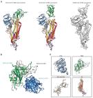
            Cryo-EM structure of the 2019-nCoV spike in the prefusion conformation

            Structure of the nCoV trimeric spike The World Health Organization has declared the outbreak of a novel coronavirus (2019-nCoV) to be a public health emergency of international concern. The virus binds to host cells through its trimeric spike glycoprotein, making this protein a key target for potential therapies and diagnostics. Wrapp et al. determined a 3.5-angstrom-resolution structure of the 2019-nCoV trimeric spike protein by cryo–electron microscopy. Using biophysical assays, the authors show that this protein binds at least 10 times more tightly than the corresponding spike protein of severe acute respiratory syndrome (SARS)–CoV to their common host cell receptor. They also tested three antibodies known to bind to the SARS-CoV spike protein but did not detect binding to the 2019-nCoV spike protein. These studies provide valuable information to guide the development of medical counter-measures for 2019-nCoV. Science, this issue p. 1260
              Bookmark
              • Record: found
              • Abstract: found
              • Article: not found

              Virological assessment of hospitalized patients with COVID-2019

              Coronavirus disease 2019 (COVID-19) is an acute infection of the respiratory tract that emerged in late 20191,2. Initial outbreaks in China involved 13.8% of cases with severe courses, and 6.1% of cases with critical courses3. This severe presentation may result from the virus using a virus receptor that is expressed predominantly in the lung2,4; the same receptor tropism is thought to have determined the pathogenicity-but also aided in the control-of severe acute respiratory syndrome (SARS) in 20035. However, there are reports of cases of COVID-19 in which the patient shows mild upper respiratory tract symptoms, which suggests the potential for pre- or oligosymptomatic transmission6-8. There is an urgent need for information on virus replication, immunity and infectivity in specific sites of the body. Here we report a detailed virological analysis of nine cases of COVID-19 that provides proof of active virus replication in tissues of the upper respiratory tract. Pharyngeal virus shedding was very high during the first week of symptoms, with a peak at 7.11 × 108 RNA copies per throat swab on day 4. Infectious virus was readily isolated from samples derived from the throat or lung, but not from stool samples-in spite of high concentrations of virus RNA. Blood and urine samples never yielded virus. Active replication in the throat was confirmed by the presence of viral replicative RNA intermediates in the throat samples. We consistently detected sequence-distinct virus populations in throat and lung samples from one patient, proving independent replication. The shedding of viral RNA from sputum outlasted the end of symptoms. Seroconversion occurred after 7 days in 50% of patients (and by day 14 in all patients), but was not followed by a rapid decline in viral load. COVID-19 can present as a mild illness of the upper respiratory tract. The confirmation of active virus replication in the upper respiratory tract has implications for the containment of COVID-19.
                Bookmark

                Author and article information

                Journal
                N Engl J Med
                N. Engl. J. Med
                nejm
                The New England Journal of Medicine
                Massachusetts Medical Society
                0028-4793
                1533-4406
                28 July 2020
                : NEJMoa2024671
                Affiliations
                From the Vaccine Research Center (K.S.C., B. Flynn, K.E.F., J.R.F., S.B.-B., A.P.W., B. Flach, S. O’Connell, A.T.N., N.D., M.M.D., N.N.N., G.S.A., D.R.F., E.L., N.A.D.-R., B.C.L., M.K.L., S. O’Dell, S.D.S., E.P., L.A.C., C.Y., J.-P.M.T., W.S., Y.Z., O.M.A., L.W., A.P., E.S.Y., K.L., T.Z., I.-T.T., A.W., I.G., L.N., R.A.G., R.J.L., J.I.M., W.-P.K., K.M.M., T.J.R., J.E.L., M.R.G., P.D.K., J.R.M., A.M., N.J.S., M.R., R.A.S., B.S.G.), the Infectious Disease Pathogenesis Section (K.W.B., M.M., B.M.N., M.G.L.), and the Biostatistics Research Branch, Division of Clinical Research (M.C.N.), National Institute of Allergy and Infectious Diseases, National Institutes of Health, Bethesda, and Bioqual (H.A., L.P., A.V.R., S.B., J.G., T.P.-T., A.S., T.-A.C., A. Cook, A.D., K.S., I.N.M.) and the Public Health Service Commissioned Corps (M.R.G.), Rockville — both in Maryland; the Department of Epidemiology, University of North Carolina at Chapel Hill, Chapel Hill (D.R.M., R.S.B.); Moderna, Cambridge, MA (D.K.E., G.S.-J., S.H., A. Carfi); and the Institute for Biomedical Sciences, George Washington University, Washington, DC (E.P.).
                Author notes
                Address reprint requests to Dr. Seder or Dr. Graham at the Vaccine Research Center, National Institute of Allergy and Infectious Diseases, National Institutes of Health, 40 Convent Drive, Bethesda, MD 20892, or at rseder@ 123456mail.nih.gov or bgraham@ 123456nih.gov .

                Drs. Seder and Graham contributed equally to this article.

                Author information
                http://orcid.org/0000-0003-1103-9608
                http://orcid.org/0000-0002-8390-1933
                http://orcid.org/0000-0002-5731-3054
                http://orcid.org/0000-0002-3287-3987
                http://orcid.org/0000-0002-1240-9044
                http://orcid.org/0000-0001-6922-6786
                http://orcid.org/0000-0003-3564-6453
                http://orcid.org/0000-0002-3935-4637
                http://orcid.org/0000-0002-7954-9674
                http://orcid.org/0000-0002-0110-881X
                http://orcid.org/0000-0002-3743-5281
                Article
                NJ202007283830001
                10.1056/NEJMoa2024671
                7449230
                32722908
                2793d25f-f957-4408-b85e-7d3224140e9d
                Copyright © 2020 Massachusetts Medical Society. All rights reserved.

                This article is made available via the PMC Open Access Subset for unrestricted re-use, except commercial resale, and analyses in any form or by any means with acknowledgment of the original source. These permissions are granted for the duration of the Covid-19 pandemic or until revoked in writing. Upon expiration of these permissions, PMC is granted a license to make this article available via PMC and Europe PMC, subject to existing copyright protections.

                History
                Categories
                Original Article
                Custom metadata
                2020-07-28T15:00:00-04:00
                2020
                07
                28
                15
                00
                00
                -04:00

                Comments

                Comment on this article